
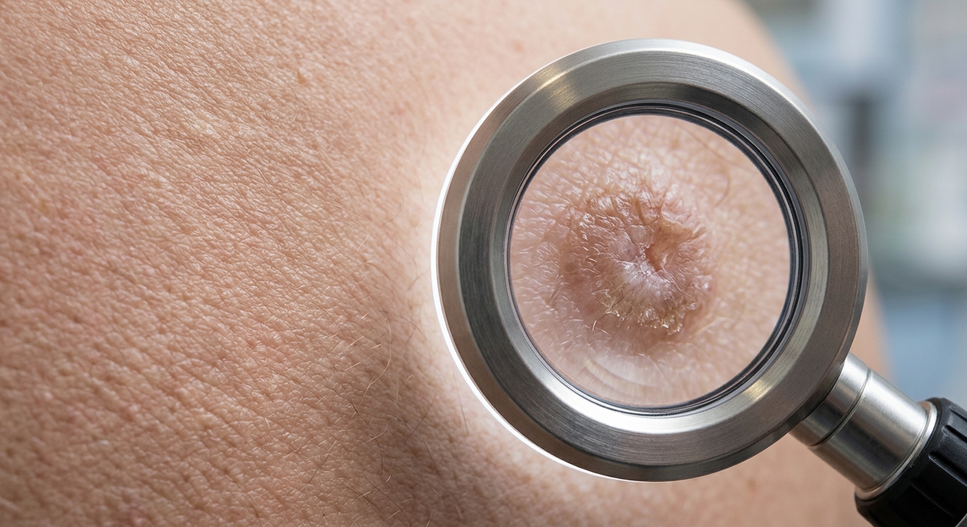
Verruca vulgaris (common warts) are benign epidermal hyperplasias caused by infection with non-oncogenic strains of Human Papillomavirus (HPV). Transmitted via direct skin contact or autoinoculation, clinical management relies on distinguishing non-malignant viral subtypes, recognizing classic lesion morphology, and implementing targeted destructive or immunomodulatory therapies when conservative watch-and-wait approaches are insufficient.
Core Virology & Transmission Principles
- Viral Subtypes & Non-Malignant Nature: Cutaneous warts are caused by non-oncogenic HPV strains (primarily HPV-1, 2, 3, 4, 7, and 10). Unlike mucosal high-risk strains (e.g., HPV-16, 18), cutaneous HPV strains do not carry malignant potential.
- Pathophysiology: Viral inoculation occurs through minor epithelial trauma into basal keratinocytes, leading to localized epidermal proliferation, hyperkeratosis, acanthosis, and papillomatosis.
- Transmission Dynamics: Spreads via direct person-to-person contact, autoinoculation (e.g., scratching, shaving), or fomites (e.g., public showers, pool decks).
- Watch-and-Wait Strategy: Because host cell-mediated immunity eventually clears the virus, approximately 65–70% of cutaneous warts spontaneously resolve within 12 to 24 months without scarring. Observation is a preferred option for asymptomatic, non-disabling lesions.
1. Clinical Subtypes & Morphologic Presentations
| Wart Subtype | Associated HPV Strains | Predilection Sites | Clinical Appearance & Key Signs |
|---|---|---|---|
| Common Warts (Verruca Vulgaris) |
HPV-2, HPV-7 (also HPV-1, 4) |
Hands, fingers, periungual regions, elbows, and knees. | Firm, hyperkeratotic papules (1 mm to 1 cm) with rough, irregular (“verrucous”) surfaces. Contain dark pinpoint specks representing thrombosed capillaries. |
| Plantar Warts (Verruca Plantaris) |
HPV-1, HPV-2, HPV-4 | Plantar surface of feet, heels, and pressure points. | Thickened, hyperkeratotic endophytic lesions pushed inward by body weight. Interrupts normal dermatoglyphics (skin lines); tender with lateral compression. |
| Flat Warts (Verruca Plana) |
HPV-3, HPV-10 | Face, dorsum of hands, shins, and shaved areas. | Smooth or slightly hyperkeratotic, flat-topped, flesh-colored or light brown papules (1–5 mm). Frequently coalesce into linear clusters along scratch lines (Koebner phenomenon). |
2. Therapeutic Modalities & Management Protocols
| Therapy Class | Agent / Modality | Instructions & Clinical Notes |
|---|---|---|
| Destructive Physical Therapy | Cryotherapy (Liquid Nitrogen) | Apply liquid nitrogen to achieve a thaw time of 20–45 seconds. Administer 2 freeze-thaw cycles every 2–4 weeks until resolution. Highly effective with good cosmetic outcome, but painful; carries risk of post-inflammatory hypopigmentation. |
| First-Line Topical Keratolyics | Salicylic Acid (17% to 40% formulations) & Cantharidin® | Salicylic Acid: Daily home application after paring down hyperkeratotic tissue with a pumice stone. For plantar warts, apply 40% salicylic acid plasters every 5 days. Well-tolerated; requires weeks to months of patient adherence. Cantharidin: Vesicant that induces epidermal blistering. |
| Topical Retinoids & Irritants | Tretinoin (Retinoic Acid) & Podofilox | Tretinoin: Applied BID for 4–6 weeks; preferred treatment for flat warts on the face to avoid scarring. Podofilox / Podophyllum Resin: Applied 3 times weekly for 4–6 weeks; common skin irritation. |
| Topical Immunomodulation | Imiquimod 5% (Aldara®) | Stimulates localized interferon-α and cytokine production. Applied 3x/week at night. Excellent non-destructive option for flat or recalcitrant warts with low recurrence rates. |
| Refractory & Procedural Options | Pulsed-Dye Laser (PDL) & Intralesional Antigen Injection | Targeted laser ablation destroys vascular supply (4–6 weeks granulation). Intralesional Candida antigen injections induce systemic cell-mediated immunity for widespread recalcitrant lesions. Surgical excision is rarely indicated due to high risk of painful scarring. |
Diagnostic Differentiation & Non-Wart Mimics
Diagnosis is primarily clinical. Biopsy is indicated only for atypical, rapidly growing, or treatment-refractory lesions to exclude malignancy.
- Diagnostic Physical Sign (Thrombosed Capillaries): Paring down the hyperkeratotic surface of a wart reveals pinpoint black dots (thrombosed capillary loops). This confirms a wart and rules out a callus.
- Skin Lines (Dermatoglyphics): Warts disrupt normal skin lines, whereas calluses and corns maintain continuous skin lines across the hyperkeratotic tissue.
- Plantar Pain Sign: Plantar warts are characteristically more painful with side-to-side (lateral) squeezing, whereas corn/callus lesions are more painful with direct vertical pressure.
- Differential Rule-Outs:
- Verrucous Carcinoma (Buschke-Löwenstein Tumor): Low-grade, slow-growing squamous cell carcinoma that mimics large, giant plantar or genital warts. Requires biopsy.
- Molluscum Contagiosum: Smooth, dome-shaped papules with central umbilication caused by Poxvirus (lacks hyperkeratosis).
- Corns / Calluses: Non-infectious hyperkeratosis secondary to friction; lacks pinpoint thrombosed capillaries.
- Seborrheic / Actinic Keratoses & SCC: Must be ruled out in older adults or sun-exposed areas.
High-Yield Exam & Practice Pearls
- Plantar Wart vs. Callus Key Differentiator: Pare down the surface layer—pinpoint black dots (thrombosed capillaries) and disrupted dermatoglyphics confirm a plantar wart; intact skin lines without bleeding confirm a callus.
- Excision Avoidance Rule: Surgical excision of plantar warts is strongly discouraged because it carries a high risk of producing painful permanent scar tissue on weight-bearing surfaces.
- Koebner Phenomenon in Flat Warts: Flat warts on the face or shins often spread in a linear distribution due to autoinoculation from shaving or scratching.
- Facial Flat Wart Strategy: Avoid destructive cryotherapy or strong acids on the face due to pigmentary changes and scarring risk; prefer non-destructive topical Tretinoin or Imiquimod.
