
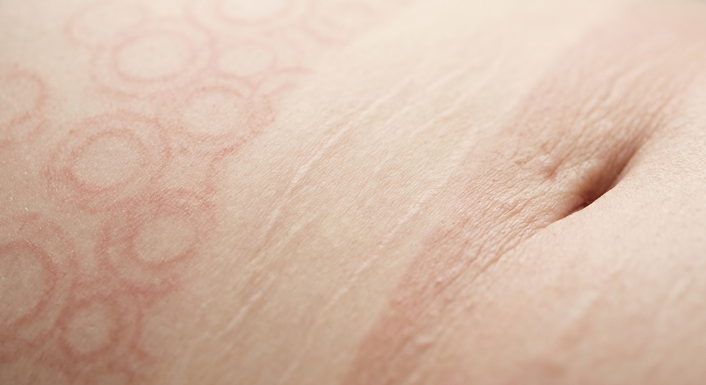
Dermatological terminology can often feel like a distinct language. Whether evaluating patients as a medical professional, studying health sciences, or reviewing clinical diagnoses, systematic classification of skin lesions is the cornerstone of accurate identification. Lesions are categorized into Primary Lesions (initial physical manifestations), Secondary Lesions (evolutionary, traumatic, or healing changes), and Morphological Patterns (spatial arrangements across the body).
Morphological Quick Reference
| Primary Lesions | Direct result of the initial disease process; key physical baseline for diagnostic differentiation. |
| Secondary Lesions | Evolve from primary lesions due to infection, physical trauma (e.g., excoriation), or the natural healing continuum. |
| Arrangement / Patterns | Distribution spatial characteristics providing crucial systemic or localized etiology clues. |
1. Primary Skin Lesions
Primary lesions reflect original structural, color, or physical changes in the skin prior to secondary alteration or evolution.
| Lesion Type | Clinical Description | Classic Example |
|---|---|---|
| Macule | Flat, non-palpable discoloration, typically <1 cm in diameter. | Freckle |
| Patch | Flat, non-palpable area of skin discoloration, ≥1 cm in diameter. | Vitiligo |
| Papule | Solid, raised lesion <1 cm; variable coloration relative to surrounding skin. | Raised nevus (mole) |
| Plaque | Elevated, plateau-like lesion ≥1 cm; often formed by coalescing papules. | Psoriasis |
| Vesicle | Fluid-filled, circumscribed elevation <1 cm in diameter. | Varicella (Chickenpox) |
| Bulla | Fluid-filled, circumscribed elevation ≥1 cm in diameter. | Second-degree burn blister |
| Pustule | Vesicle-like elevation filled with purulent exudate (pus). | Impetigo, acne |
| Nodule | Solid, palpable, elevated lesion ≥1 cm, typically extending into deep dermis or subcutis. | Epidermal cyst |
| Wheal | Transient, circumscribed area of cutaneous edema (swelling). | Hive (Urticaria) |
| Purpura | Non-blanching lesions caused by extravasated red blood cells trapped in skin tissue. | Petechiae, ecchymosis |
2. Secondary Skin Lesions
Secondary lesions develop over time as a consequence of external manipulation, superinfection, or the natural evolutionary path of a primary lesion.
| Lesion Type | Clinical Description | Classic Example |
|---|---|---|
| Excoriation | Superficial linear or punctate marks produced by physical scratching or trauma. | Pruritic dermatoses |
| Lichenification | Epidermal thickening with accentuated skin markings, secondary to chronic rubbing. | Lichen simplex chronicus |
| Fissure | Linear crack extending through the epidermis into the underlying dermis. | Split lip, tinea pedis |
| Erosion | Partial loss of the epidermis; heals without scarring. | Ruptured bullous lesion |
| Ulcer | Full-thickness loss extending through epidermis and dermis; heals with scarring. | Pressure ulcer |
| Scale | Desquamated, elevated dead epidermal cells (keratin flakes). | Dandruff, psoriasis |
| Atrophy | Thinning of epidermis/dermis leading to loss of normal skin markings and transparency. | Topical corticosteroid overuse |
3. Spatial Patterns & Distribution Configurations
Beyond individual lesion traits, evaluating spatial arrangement and body distribution provides critical clues for establishing differential diagnoses.
Common Lesion Pattern Formations
- Annular: Ring-shaped formation with central clearing (e.g., Erythema migrans in Lyme disease, Tinea corporis).
- Confluent / Coalescent: Multiple individual lesions merging into larger, continuous clusters or plaques.
- Reticular: Net-like, lacy, or reticulated distribution pattern across the cutaneous surface.
- Dermatomal: Unilateral distribution confined strictly along a neurocutaneous sensory dermatome (e.g., Herpes zoster / Shingles).
- Linear: Formed in distinct straight lines or linear streaks, often indicating exogenous exposure (e.g., Rhus dermatitis from poison ivy).
Clinical Practice Pearls
- Perform Systematic Palpation: Distinguish macules from papules and patches from plaques through tactile inspection rather than visual assessment alone.
- Assess Blanching Capacity: Apply direct pressure to reddish-purple lesions to differentiate vascular dilation (blanchable) from extravasated red cells in purpura (non-blanching).
- Trace Distribution Patterns: Identifying a linear or dermatomal configuration often narrows differential diagnostic possibilities immediately.
