
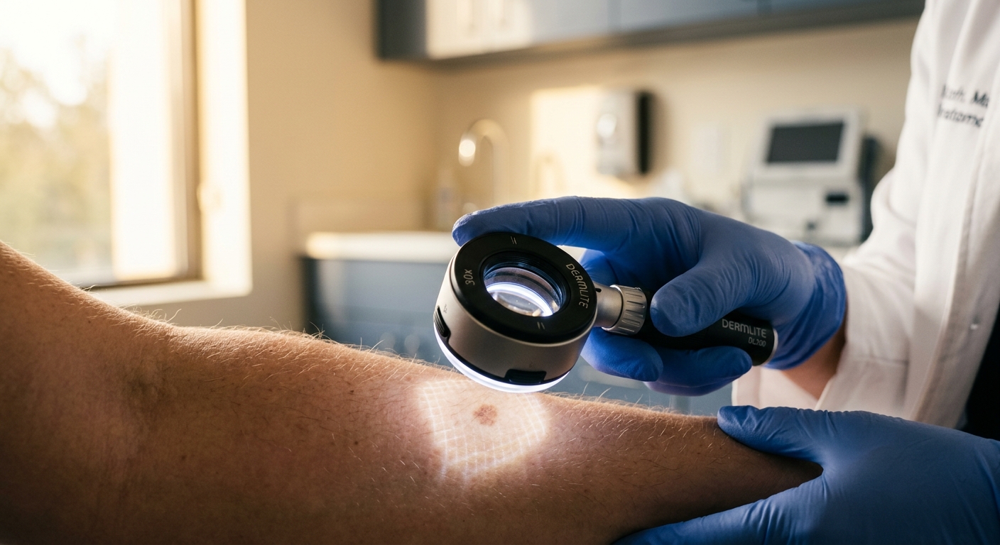
Skin cancer encompasses precancerous dysplasias, non-melanoma skin cancers (NMSC), and cutaneous malignant melanoma. Early clinical identification through diagnostic mnemonics, prompt histological biopsy confirmation, and appropriate surgical or topical interventions are critical to preventing severe local destruction and systemic metastasis.
Core Epidemiology & Ultraviolet Risk Factors
- Ultraviolet Radiation Exposure: Chronic cumulative UV exposure drives non-melanoma skin cancers (BCC and SCC). Intense, intermittent blistering sunburns—especially ≥5 sunburns before age 20—increase malignant melanoma risk by 80%.
- Sunscreen Nuance: Consistent high-SPF sunscreen significantly reduces the incidence of Basal Cell Carcinoma and Squamous Cell Carcinoma, but shows a less direct effect in mitigating melanoma risk (which is more tied to genetic predisposition and intense sporadic exposure).
- Precancerous Evolution: Actinic Keratoses (AK) represent UV-induced intraepidermal dysplasias with a ~1% annual transformation rate into invasive Squamous Cell Carcinoma.
- Biopsy Mandate: Any changing, suspicious, or non-healing cutaneous lesion requires definitive tissue biopsy prior to definitive surgical or destructive management.
1. Clinical Identification & Diagnostic Mnemonics
| Malignancy / Lesion Type | Diagnostic Mnemonic | Key Features & Clinical Presentation |
|---|---|---|
| Malignant Melanoma (Melanocyte Origin) |
A-B-C-D-E Rule |
A – Asymmetric morphology. B – Borders (irregular, notched, or blurred). C – Color variation (shades of black, brown, red, white, or blue). D – Diameter (>6 mm; pencil eraser size). E – Evolving (new, enlarging, or changing lesions; most critical indicator). |
| Basal Cell Carcinoma (BCC) (Basal Layer Origin) |
P-U-T O-N |
P – Pearly papule with rolled margins. U – Ulcerating center (“rodent ulcer”). T – Telangiectasias (arborizing blood vessels). O – On sun-exposed areas (face, scalp, pinnae). N – Nodular morphology (slow-growing, low metastatic potential). |
| Squamous Cell Carcinoma (SCC) (Keratinocyte Origin) |
N-O S-U-N |
N – Nodular/hyperkeratotic. O – Opaque surface with thick scaling/crusting. S – Sun-exposed areas (lower lip, ears, dorsum of hands). U – Ulcerating base with firm elevated borders. N – Non-distinct margins (faster growth; low but real risk of metastasis). |
| Actinic Keratosis (AK) (Precancerous Precursor) |
“Sandpaper Touch” | Small (3–10 mm), rough, erythematous scaly patches. Often felt better than seen on light palpation. Recurrent crusting occurs after picking. Precursor to invasive SCC. |
2. Therapeutic Modalities & Management Protocols
| Indication | Therapy Class / Agent | Regimen & Practice Considerations |
|---|---|---|
| Actinic Keratoses (AK) (Field vs. Targeted Therapy) |
Cryotherapy (Liquid Nitrogen) | Targeted destructive therapy for isolated lesions. Causes localized blistering/crusting (~2 weeks) with excellent cosmetic outcomes. |
| Topical Chemotherapy 5% Fluorouracil (5-FU) |
Daily Regimen: Applied 1–2x daily for 2–4 weeks (causes intense inflammatory reaction and crusting). Pulse Regimen: Applied 1–2 days/week for 7–10 weeks (provides comparable field efficacy with reduced discomfort). |
|
| Alternative Field Topicals Imiquimod, Diclofenac, Ingenol mebutate, PDT |
Imiquimod 5%: Immune response modifier. Photodynamic Therapy (PDT): Topical δ-aminolevulinic acid followed by light activation. Ingenol mebutate (Picato®): Rapid 2-day (trunk) or 3-day (face) topical dosing. |
|
| Invasive Cutaneous Cancers (BCC & SCC) |
Mohs Micrographic Surgery | Gold standard for lesions on cosmetically/functionally sensitive areas (face, ears, hands) or tumors with aggressive histology. Provides 100% margin evaluation with maximum tissue preservation. |
| Surgical Excision & Non-Surgical Destruction | Standard excision with clean margins (4–6 mm). Non-surgical options for low-risk superficial BCC/SCC include Electrodesiccation & Curettage (ED&C), radiation therapy, or topical 5-FU/Imiquimod. | |
| Malignant Melanoma | Wide Local Excision & Staging | Requires full-thickness wide surgical excision with margin width determined by Breslow depth. Lesions >0.8–1.0 mm depth warrant Sentinel Lymph Node Biopsy (SLNB) for pathological staging. |
Diagnostic Evaluation & Biopsy Protocols
Definitive management of suspicious cutaneous lesions requires accurate histological staging.
- Full-Thickness Excisional Biopsy: The standard of care for suspected melanoma. Shave biopsies that truncate the base are contraindicated because they impede accurate determination of Breslow depth (the single most important prognostic factor).
- Punch or Shave Biopsy: Appropriate for suspected BCC, SCC, or AK when complete excision is not initially feasible.
- Dermoscopy: Non-invasive evaluation revealing pigment network alterations, streak structures, blue-white veils (melanoma), or arborizing telangiectasias (BCC).
High-Yield Exam & Practice Pearls
- Melanoma Prognostic Metric: Breslow thickness (measured in millimeters from the stratum granulosum to the deepest tumor cell) is the single most crucial determinant of melanoma staging and overall prognosis.
- Biopsy Choice Pitfall: Never perform a superficial shave biopsy or cryoablation on a lesion suspected of being malignant melanoma. Always obtain a full-thickness excisional biopsy with narrow margins.
- Marjolin’s Ulcer: Aggressive Squamous Cell Carcinoma arising within chronic non-healing wounds, burn scars, or osteomyelitis sinus tracts. Carries a significantly higher risk of metastasis.
- Keratoacanthoma: Rapidly growing, dome-shaped papule with a central keratin-filled plug. Considered a low-grade subtype of SCC; treated with complete surgical excision due to potential for rapid growth.
- Lower Lip Lesion Risk: SCC occurring on sun-exposed mucosal surfaces (such as the lower lip or pinna of the ear) has a significantly higher metastatic rate than cutaneous SCC elsewhere on the body.
