
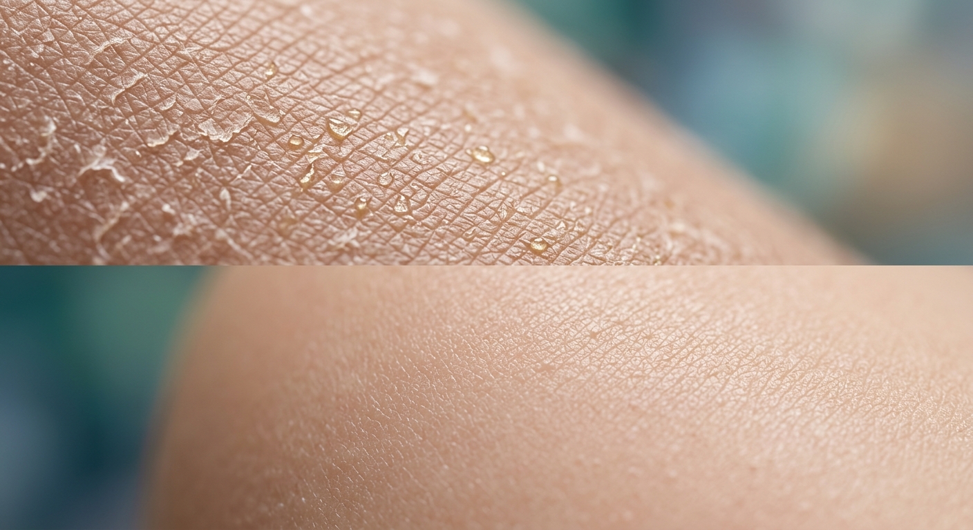
Seborrheic dermatitis is a common, chronic-relapsing inflammatory skin disease affecting sebum-rich areas of the head, face, and trunk. Driven primarily by an abnormal inflammatory immune response to commensal Malassezia yeasts, management centers on topical antifungal agents, mild anti-inflammatory therapies, and keratolytic scaling control.
Core Pathophysiology & Disease Associations
- Malassezia Hypothesized Etiology: Lipophilic yeasts (predominantly Malassezia globosa and M. restricta) metabolize sebum triglycerides into free fatty acids, triggering epidermal inflammation, alternative complement activation, and barrier dysfunction in susceptible hosts.
- Anatomic Sebum Predilection: Confined strictly to sebaceous gland-rich regions: scalp (dandruff/pityriasis capitis), eyebrows, glabella, eyelid margins (blepharitis), nasolabial folds, retroauricular skin, external ear canals, and pre-sternal chest.
- Neurologic & Systemic Triggers: Unusually severe, widespread, or recalcitrant disease is strongly associated with Parkinson’s disease (due to hyperseborrhea), HIV/AIDS (low CD4 counts), and acute central nervous system trauma/stroke.
- Infantile Form (“Cradle Cap”): Self-limiting eruption in infants (<3 months) manifesting as thick, greasy, yellow crusts on the scalp. Unlike diaper dermatitis or eczema, it typically spares the diaper region and causes minimal pruritus.
1. Clinical Manifestations & Differential Considerations
| Patient Demographics | Anatomic & Pathologic Features | Clinical Appearance |
|---|---|---|
| Infants (0–3 months) |
Scalp, vertex, retroauricular folds, and facial folds. Diaper region is typically spared. | Adherent, greasy, yellowish scaly crusts over erythematous skin (“cradle cap”). Generally asymptomatic without distress or severe itch. |
| Adolescents & Adults (Post-puberty) |
Scalp, eyebrows, glabella, nasolabial folds, moustache area, chest, and upper back. | Erythematous plaques covered with greasy, yellowish, or salmon-colored scales. Waxing and waning course; frequently worsens in winter. |
| Ophthalmic Variant | Eyelid margins, meibomian glands, and eyelashes. | Blepharitis with anterior eyelid margin erythema, crusting along lash lines, conjunctival injection, and burning sensation. |
| High-Risk Populations (HIV / Parkinson’s) |
Generalized distribution; face, neck, trunk, and intertriginous areas. | Extremely severe, explosive, extensive erythematous scaly plaques resistant to standard topical therapies. |
2. Therapeutic Classes & Evidence-Based Options
| Therapy Class | Medications / Options | Clinical Rationale & Practice Considerations |
|---|---|---|
| First-Line Topical Antifungals | Ketoconazole 2% cream/shampoo, Ciclopirox 1%, Selenium Sulfide 2.5%, Zinc Pyrithione | Directly reduces Malassezia density. Shampoos should be left on the scalp for 5–10 minutes before rinsing, used 2–3 times weekly. First-line therapy for both scalp and body. |
| Anti-Inflammatory Modalities | Low-potency corticosteroids (Class IV-VII e.g., Hydrocortisone 1%, Desonide), Topical Calcineurin Inhibitors (Tacrolimus, Pimecrolimus) | Used short-term for acute inflammatory flares. Low-potency topical steroids minimize risks of skin atrophy and telangiectasia on the face. Topical calcineurin inhibitors are ideal non-steroidal maintenance agents for facial folds. |
| Keratolytic Agents | Salicylic acid, Coal tar preparations, Sulfur/Sulfonamide combinations | Helps soften and shed thick, adherent scales. Coal tar slows epidermal proliferation but carries odor/staining drawbacks. Mineral oil or petroleum jelly can soften infantile scalp crusts prior to gentle combing. |
| Systemic Antifungals | Oral Ketoconazole, Fluconazole, Itraconazole | Reserved strictly for severe, recalcitrant, widespread disease or underlying immunocompromise failing all topical regimens. Monitor baseline LFTs. |
Diagnostic Evaluation & Differential Workup
Diagnosis is clinical, based on lesion morphology and distribution. Diagnostic testing is rarely required except to exclude key mimics.
- Clinical Diagnosis: Characterized by yellowish, greasy scales on erythematous bases in sebaceous distributions.
- Fungal Culture / KOH Prep: Useful in pediatric patients to rule out Tinea Capitis (which displays alopecia, broken hairs, and posterior cervical lymphadenopathy—features absent in seborrhea).
- Differential Differentiation:
- Psoriasis Vulgaris: Features thick, micaceous silvery scales on extensor surfaces, sharply demarcated borders, and nail pitting (versus indistinct, greasy yellowish scales on flexor/facial folds in seborrhea).
- Atopic Dermatitis: Intensely pruritic, poorly demarcated papules/plaques on flexor surfaces; spares the nasolabial folds and scalp vertex in adults.
- Rosacea: Erythema and telangiectasias over the central face with papules/pustules; lacks the greasy scaling of seborrheic dermatitis.
High-Yield Exam & Practice Pearls
- Systemic Association Red Flags: Sudden-onset, severe, or explosive seborrheic dermatitis should prompt clinical screening for HIV infection or Parkinson’s disease.
- “Cradle Cap” Management Rule: In infants, avoid strong medicated shampoos or topical steroids. Recommend mild baby shampoo, application of warm mineral/olive oil to soften crusts, and gentle brushing with a soft toothbrush.
- Facial Steroid Avoidance: High-potency topical corticosteroids must never be used on the face due to rapid development of steroid-induced rosacea, skin atrophy, telangiectasias, and rebound flares.
- Tinea Capitis Key Differentiator: Hair loss (alopecia) and tender cervical/occipital lymphadenopathy favor Tinea Capitis over Seborrheic Dermatitis.
- Biologic Contraindication: Systemic biologic agents (e.g., TNF or Interleukin inhibitors) are not indicated for seborrheic dermatitis.
