
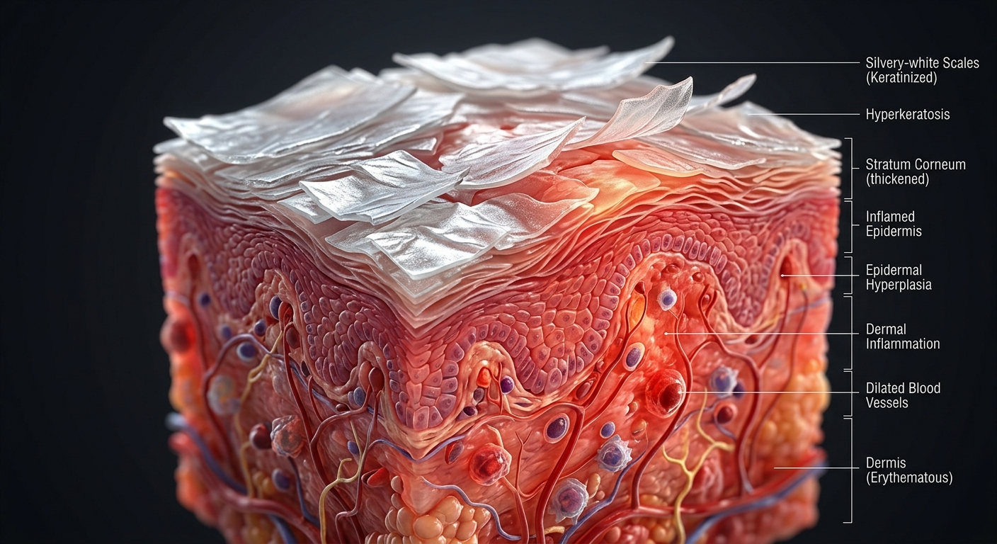
Psoriasis vulgaris is a chronic, immune-mediated inflammatory skin disorder driven by T-cell-mediated hyperproliferation of keratinocytes and incomplete cell maturation. Characterized by well-demarcated erythematous plaques covered with silvery scales, effective long-term management depends on disease severity stratification, screening for joint involvement, and stepwise therapy from topicals to targeted biologic agents.
Core Pathophysiology & Severity Classification
- Pathophysiology: Dendritic cell activation releases IL-12 and IL-23, activating Th17 and Th1 cells. Overproduction of IL-17, IL-22, and TNF-α causes rapid epidermal transit time (3–5 days vs. normal 28–30 days), leading to incomplete keratinocyte maturation, acanthosis, and hyperkeratosis.
- Triggers & Risk Factors: Genetic predisposition (HLA-Cw6) combined with environmental triggers including mechanical trauma (Koebner phenomenon), stress, cold weather, and drugs (beta-blockers, lithium, antimalarials, systemic steroid withdrawal).
- Severity Stratification by Body Surface Area (BSA):
- Mild: <2% BSA (easily controlled with targeted topical agents).
- Moderate: 3% to 10% BSA (may require phototherapy or systemic therapy).
- Severe: >10% BSA (requires systemic or biologic therapies; immediate dermatology referral).
1. Clinical Presentation & Subtypes
| Psoriasis Subtype | Anatomic & Pathologic Features | Clinical Appearance & Associations |
|---|---|---|
| Plaque Psoriasis (Psoriasis Vulgaris) |
Symmetric involvement of extensor surfaces (knees, elbows), scalp, umbilicus, and lumbosacral region. | Most common form (>80–90%). Well-demarcated, salmon-pink erythematous plaques with overlying loose, micaceous silvery scales. Positive Auspitz sign. |
| Guttate Psoriasis | Mainly affects the trunk and proximal extremities; abrupt onset. | “Dewdrop-like” small (0.5–1.5 cm) erythematous papules with fine scale. Characteristically develops 2 to 3 weeks after Group A Streptococcal pharyngitis. |
| Inverse (Intertriginous) | Affects skin folds: axillae, groin, inframammary, and intergluteal cleft. | Smooth, shiny, deep red erythematous plaques lacking silvery scale due to friction and moisture in skin folds. |
| Erythrodermic & Pustular | Generalized involvement (>90% BSA) or widespread sterile pustules (von Zumbusch type). | Dermatologic emergencies requiring hospitalization. High risk for hypothermia, high-output heart failure, electrolyte imbalances, and sepsis. |
| Psoriatic Arthritis (PsA) | Inflammatory arthropathy affecting peripheral joints and axial skeleton. | Occurs in ~20% of psoriasis patients. Key signs include dactylitis (“sausage digits”), enthesitis (Achilles tendonitis), and nail dystrophy. |
2. Diagnostic Evaluation & Workup
| Diagnostic Modality | Typical Findings | Clinical Utility & Differential Considerations |
|---|---|---|
| Physical Signs & Examination | Auspitz sign & Koebner phenomenon | Auspitz Sign: Pinpoint bleeding upon forcible scraping of scale due to elongated dermal papillae. Koebner Phenomenon: Development of new psoriatic lesions along lines of skin trauma. |
| Serology & Inflammatory Markers | RF Negative, anti-CCP Negative, normal/elevated ESR/CRP | Helps differentiate Psoriatic Arthritis (a seronegative spondyloarthropathy) from Rheumatoid Arthritis. Hyperuricemia may occur due to high epidermal cell turnover. |
| Radiographs (Joints) | “Pencil-in-cup” deformity & DIP joint space narrowing | Classic radiologic sign of advanced PsA in distal interphalangeal (DIP) joints showing osteolysis of the proximal phalanx telescoping into the distal unit. |
| Histopathology (Biopsy) | Acanthosis, hyperkeratosis, parakeratosis, Munro microabscesses | Reserved for atypical cases. Shows regular epidermal hyperplasia, loss of stratum granulosum, and neutrophil collection in the stratum corneum (Munro microabscesses). |
Stepwise Treatment Modalities & Adverse Effect Profiles
Therapy choice is driven by BSA involvement, anatomic site, and systemic/joint manifestations.
- Mild Disease (<2–3% BSA) — Topicals First-Line:
- Topical Corticosteroids: High-potency (Clobetasol) for body plaques; low-potency (Hydrocortisone) for face/intertriginous areas. Limit high-potency use to <2–4 weeks to avoid atrophy, striae, and tachyphylaxis.
- Vitamin D3 Analogues (Calcipotriene): Inhibits keratinocyte proliferation. Commonly combined with topical corticosteroids (e.g., Taclonex®) for synergistic efficacy.
- Second-Line Topicals: Tazarotene (topical retinoid), Anthralin, and Coal Tar preparations (useful for scalp psoriasis).
- Moderate-to-Severe Disease (>10% BSA) or Refractory Cases:
- Phototherapy (Narrowband UVB / UVA): Highly effective for generalized plaque psoriasis (>30% BSA); suppresses DNA synthesis and immune response.
- Oral Systemic Non-Biologics: Methotrexate (inhibits DHFR; requires LFT & CBC monitoring), Cyclosporine (rapid control; risk of nephrotoxicity and hypertension), Acitretin (oral retinoid; highly teratogenic, avoid pregnancy for 3 years post-use), and Apremilast (oral PDE-4 inhibitor).
- Biologic Targeted Therapies:
- TNF-α Inhibitors: Adalimumab, Etanercept, Infliximab.
- IL-12/23 & IL-23 Inhibitors: Ustekinumab, Guselkumab, Risankizumab.
- IL-17 Inhibitors: Secukinumab, Ixekizumab, Brodalumab.
3. Differential Diagnosis & Non-Psoriatic Lookalikes
Rule-Out Conditions (Papulosquamous Mimics)
- Seborrheic Dermatitis: Greasy, yellowish scales on erythematous skin concentrated on scalp, nasolabial folds, and chest; lacks the thick, micaceous silver scale of psoriasis.
- Pityriasis Rosea: Starts with an initial “herald patch” followed 1–2 weeks later by a self-limiting “Christmas tree” distribution of oval, salmon-pink macules along skin cleavage lines.
- Lichen Planus: Characterized by the 6 Ps: Purple, Pruritic, Polygonal, Planar, Papules, and Plaques, often with white fine lines (Wickham striae) on flexor surfaces.
- Nummular Dermatitis: Coin-shaped, intensely pruritic vesicular/crusted plaques on extremities without classic silvery scaling or extensor predilection.
- Cutaneous Squamous Cell Carcinoma (sCC) / Bowen’s Disease: Solitary, non-healing, persistent scaly or crusted red plaque; warrants biopsy if unresponsive to topical steroids.
High-Yield Exam & Practice Pearls
- Systemic Steroid Warning: Never administer oral systemic corticosteroids (e.g., Prednisone) for plaque psoriasis. Sudden withdrawal can precipitate severe, life-threatening generalized pustular or erythrodermic psoriasis rebound.
- Pre-Biologic Screening Protocol: Prior to initiating TNF-α or Interleukin inhibitors, screen all patients for latent Tuberculosis (PPD/QuantiFERON-TB Gold) and Hepatitis B/C due to severe reactivation risks.
- Streptococcal Connection: Always obtain a throat swab or anti-streptolysin O (ASO) titer in young patients presenting with a sudden outbreak of guttate psoriasis. Treating underlying strep can lead to resolution.
- Acitretin Pregnancy Rule: Acitretin is strictly teratogenic. Female patients of childbearing potential must avoid pregnancy during treatment and for at least 3 years after discontinuation due to prolonged lipophilic accumulation.
- Classic Radiographic Sign: “Pencil-in-cup” deformity on hand/foot X-rays is pathognomonic for advanced Psoriatic Arthritis affecting the DIP joints.
