
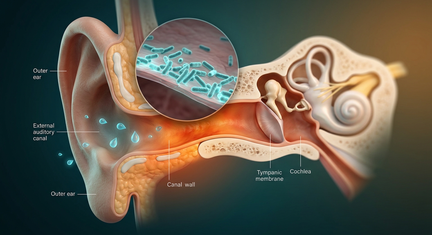
Otitis Externa (OE)—commonly referred to as “Swimmer’s Ear”—is an acute inflammation or infection of the external auditory canal (EAC), the auricle, or both. It develops when microenvironmental disruptions (excess moisture or mechanical trauma) break down the ear canal’s protective lipid barrier and acidic mantle, predisposing the tissue to opportunistic bacterial or fungal proliferation.
Etiology, Risk Factors & Pathogen Profile
- Primary Microenvironmental Triggers:
- Local Microtrauma: Vigorous use of cotton swabs (Q-tips), hearing aid usage, earplug retention, or cerumen impaction.
- Environmental Moisture: Frequent swimming or prolonged freshwater exposure (higher risk than saltwater) that washes away protective cerumen and macerates epithelium.
- Predominant Bacterial Pathogens:
- Pseudomonas aeruginosa (Most common bacterial driver; memory hook: Pool → Pseudomonas).
- Staphylococcus aureus and Staphylococcus epidermidis.
- Anaerobic co-pathogens (less common).
- Fungal Pathogens (Otomycosis): Aspergillus species (most common fungal cause, e.g., A. niger) and Candida species.
1. Clinical Presentation & Diagnostic Findings
| Clinical Variant | Characteristic Features & Exam Findings | Key Clinical Pearls & Differential |
|---|---|---|
| Classic Bacterial Otitis Externa |
• Hallmark Exam Sign: Severe ear pain reproduced by tragus palpation or traction on the pinna. • Rapid-onset otalgia (1–2 days), ear fullness, and conductive hearing loss secondary to canal edema. • Purulent, serous, or foul-smelling otorrhea. • Otoscopy: Canal erythema, marked edema, luminal narrowing, and sloughed epithelial debris. |
• Acute Otitis Media (AOM): Tragus/pinna movement is non-painful; tympanic membrane is bulging or immobile. • Contact Dermatitis: Severe pruritus without severe tenderness or canal occlusion. • Herpes Zoster Oticus (Ramsay Hunt): Facial palsy with vesicular eruptions in EAC/pinna. |
| Fungal Otitis Externa (Otomycosis) |
• Clinical Rule: Features more ITCH (pruritus) and less PAIN compared to bacterial OE. • Otoscopy reveals thick white, gray, green, or black exudate with visible hyphae/spores (often resembling “wet newspaper”). |
2. Diagnostic & Procedural Strategy
| Diagnostic Pillar | Clinical Method | Rationale & Indications |
|---|---|---|
| Primary Assessment | Clinical History & Otoscopy | Otitis Externa is fundamentally a clinical diagnosis. Diagnostic testing or routine swabs are not recommended for standard presentations because causative pathogens are highly predictable. |
| Microbiological Culture | Bacterial & Fungal Swab Culture | Reserved exclusively for treatment failure, recurrent disease, atypical presentations, or immunocompromised hosts. |
| Canal Debridement | Gentle Dry Suction / Aspiration | Removing obstructive cerumen and inflammatory debris enhances direct contact of topical drops. Irrigation is contraindicated during acute infection or if TM status is uncertain. |
| Edema Management | Otic Ear Wick Placement | Indicated when severe canal swelling prevents drop penetration. The wick acts as a conduit to transport topical drops deep into the canal; re-evaluate or remove in 2–3 days. |
3. Comprehensive Treatment & Safety Protocols
| Therapy Class | Agent / Regimen | Clinical Guidance & Safety Warnings |
|---|---|---|
| Analgesic Support | Oral NSAIDs (e.g., Ibuprofen) or Acetaminophen. | Essential for first 24–48 hours until topical antimicrobial agents significantly reduce localized canal inflammation. |
| First-Line Topical Antimicrobials |
• Fluoroquinolones: Ofloxacin 0.3% drops or Ciprofloxacin 0.2% drops. • Combination: Neomycin / Polymyxin B / Hydrocortisone. |
Topical therapy is the cornerstone of cure. Corticosteroid additions accelerate symptom relief and edema resolution. Apply for 7–10 days. |
| Acidifying Agents | Acetic Acid 2% drops. | Alters canal pH to suppress microbial growth; ideal for mild cases or prevention, though may cause transient stinging. |
| Antifungal Topicals | Clotrimazole 1% or Acetic Acid drops. | First-line for otomycosis following thorough canal debridement. |
CRITICAL SAFETY ALERT: Tympanic Membrane Integrity & Ototoxicity
If the tympanic membrane (TM) is ruptured, perforated, or cannot be visualized due to edema, NEVER use otic preparations containing Neomycin, Gentamicin, or Aminoglycosides due to risk of sensorineural ototoxicity. Use non-ototoxic Fluoroquinolone drops (Ofloxacin or Ciprofloxacin) only.
4. Severe Complication: Malignant (Necrotizing) Otitis Externa
Malignant Otitis Externa is an aggressive, life-threatening infection that extends beyond the EAC cartilage into the adjacent soft tissue, mastoid, skull base, and temporal bone (causing skull base osteomyelitis). Despite its name, it is an invasive infectious process, not a malignancy.
| Feature | Uncomplicated Otitis Externa | Malignant (Necrotizing) Otitis Externa |
|---|---|---|
| Patient Population | Swimmers, cotton swab users, general public. | Immunocompromised: Elderly diabetics (poorly controlled), HIV/AIDS, active chemotherapy. |
| Primary Pathogen | Pseudomonas aeruginosa, S. aureus. | Pseudomonas aeruginosa (>95% of cases). |
| Pain Profile | Moderate, reproduced by tragus/pinna movement. | Severe, deep nocturnal ear pain disproportionate to exam findings. |
| Complications | Local canal edema, temporary conductive hearing loss. | Temporal bone osteomyelitis, Cranial Nerve palsies (CN VII most common, followed by IX, X, XI). |
| Primary Therapy | Topical antimicrobial drops. | Systemic (IV) anti-pseudomonal antibiotics (e.g., IV Ciprofloxacin, Cefepime, or Piperacillin-tazobactam) + Urgent ENT surgical consultation. |
| Diagnostic Imaging | Not indicated. | CT scan of temporal bone (preferred for bone erosion); MRI or Technetium-99m bone scan for soft tissue/osteomyelitis tracking. |
High-Yield Exam & Practice Pearls
- The Quintessential Exam Finding: Tragus tenderness or pain with pinna traction unequivocally isolates pathology to the external ear canal.
- Classic Bacteriology: Pseudomonas aeruginosa is the primary culprit in bacterial OE.
- Fungal Signatures: Itching > Pain + thick white, gray, or black exudate points to Otomycosis.
- Ototoxic Contraindication: Non-intact TM = Avoid Neomycin/Aminoglycosides; use Fluoroquinolones (Ofloxacin/Ciprofloxacin).
- Severe Edema Workaround: Place an ear wick to ensure topical drops reach the deep EAC when swollen shut.
- Red Flag Combination: Diabetic / Immunocompromised Patient + Severe Disproportionate Nocturnal Pain = 🚨 Malignant Otitis Externa. Order CT and consult ENT immediately.
