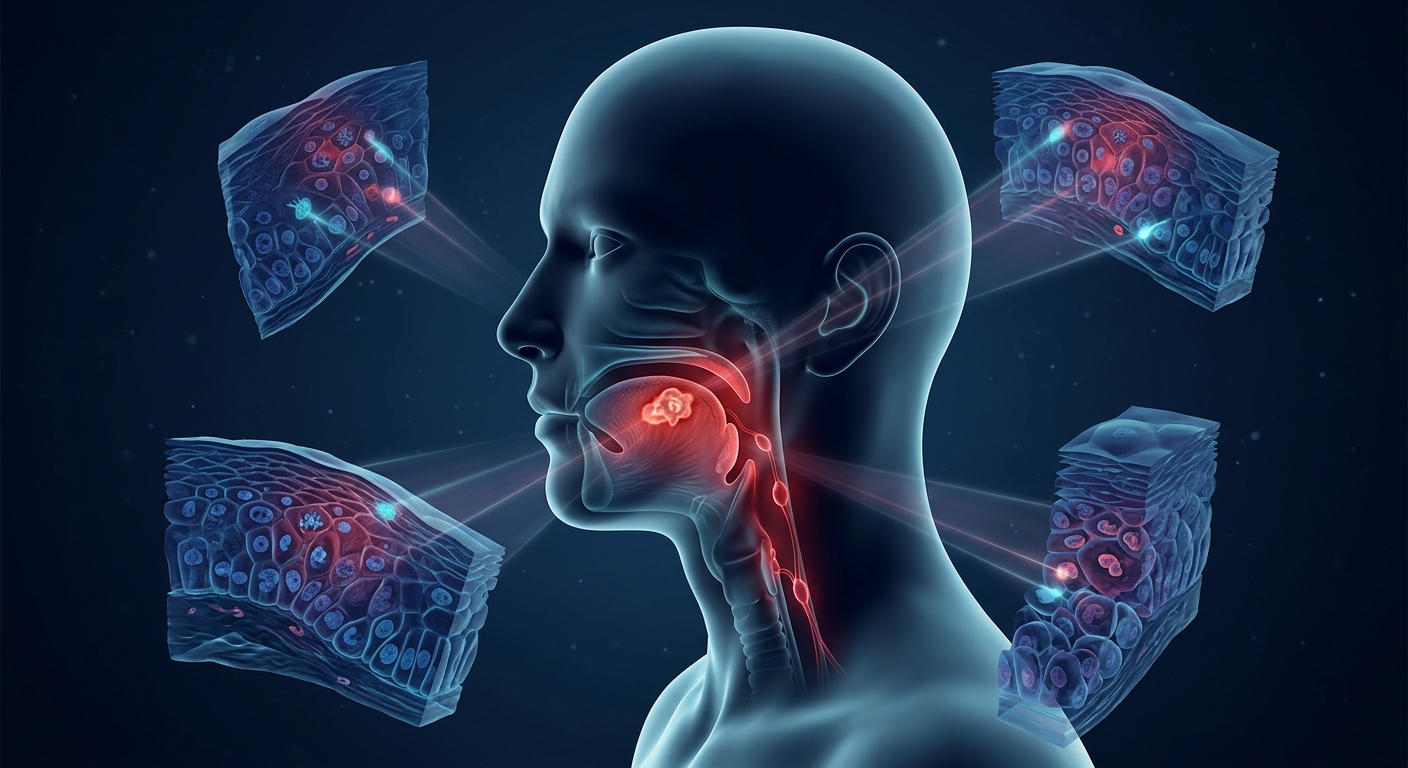
Oral Squamous Cell Carcinoma (SCC) represents the vast majority of oral cavity malignancies. Because early-stage lesions are frequently asymptomatic or non-painful, diagnosis is often delayed for months. Recognizing subtle red-flag characteristics, distinguishing malignant lesions from common benign infectious conditions, and securing prompt histological evaluation are essential for improving patient outcomes.
Epidemiology, Risk Factors & High-Risk Profile
- Histological Dominance: ~95% of all oral cavity cancers are pathologically classified as Squamous Cell Carcinoma (SCC).
- Primary Environmental Drivers:
- Tobacco Exposure: Cigarettes, cigars, pipes, and smokeless/dipping tobacco (dose-dependent risk).
- Heavy Alcohol Consumption: Acts synergistically with tobacco to multiply overall carcinogenic risk.
- High-Risk Viral Infection: HPV-16 is strongly linked to oropharyngeal and oral head and neck malignancies.
- Classic Demographic Profile: Male sex predominance; advancing age (>66% of cases diagnosed in patients aged >55 years).
1. Differential Diagnosis: Benign vs. Malignant Lesions
| Clinical Feature |
Benign / Infectious (HSV, Aphthous, Candida) |
Oral Squamous Cell Carcinoma (SCC) |
| Symptom Onset & Pain |
Acute onset, highly painful, tender, and sensitive. |
Indolent onset, typically painless in early stages. |
| Lesion Duration |
Self-limiting; resolves completely within 1–2 weeks. |
Persistent; fails to heal after weeks or months. |
| Base & Consistency |
Soft, superficial, non-indurated base. |
Firm, indurated base with tissue fixation. |
| Morphology |
Vesicles, shallow erosions, removable white plaques. |
Deep ulceration with rolled borders, leukoplakia/erythroplakia, or exophytic masses. |
2. Regional Lymphadenopathy Risk Stratification
| Lymph Node Feature |
Infectious / Reactive Etiology |
Malignant Etiology (SCC Metastasis) |
| Palpation & Tenderness |
Tender, painful to touch. |
Nontender / Asymptomatic. |
| Mobility & Texture |
Mobile, soft to firm consistency. |
Fixed / Immobile, hard/stony induration. |
| Size & Progression |
Variable size; fluctuates and resolves as infection clears. |
>1 cm, progressive enlargement over time. |
The Golden Clinical Rule: The 2-Week Rule
Any unexplained oral mucosal lesion or ulceration that fails to heal after 14 days of conservative management requires immediate referral for specialist evaluation and diagnostic tissue biopsy.
3. Diagnostic Pathways & Screening Guidelines
| Evaluation Step |
Standard Clinical Protocol |
Key Rationale & Guidelines |
| Specialist Referral |
Prompt referral to Otolaryngology (ENT) or Oral/Maxillofacial Surgery. |
Physical exam alone cannot reliably confirm or rule out malignancy. |
| Definitive Diagnostics |
Tissue Biopsy (Gold Standard). |
Provides essential histological confirmation, tumor grading, and invasion depth. |
| Screening Recommendations |
Routine dental examinations & opportunistic visual inspection/palpation. |
USPSTF concludes current evidence is insufficient for routine population-wide screening in asymptomatic adults (does not apply to symptomatic/visible lesions). |
High-Yield Exam & Practice Pearls
- Histology Majority: ~95% of oral cavity malignancies are Squamous Cell Carcinomas.
- Lifestyle Synergy: Tobacco (all forms) is the leading individual risk factor; combining tobacco and heavy alcohol creates a synergistic carcinogenic effect.
- Viral Trigger: High-risk HPV Type 16 plays a major role in head and neck SCC development.
- The Diagnostic Trap: Early oral SCC is classically painless. Lack of pain should never provide false reassurance.
- Classic Triad: Persistent (>2 weeks), firm/indurated base, and ulcerated or exophytic morphology.
- Malignant Node Profile: Cervical nodes that are >1 cm, hard, nontender, and fixed/immobile indicate regional metastasis.
- Definitive Action: Physical examination cannot establish a final diagnosis—tissue biopsy via specialist referral is required.
- Timeline Divider: Benign infectious lesions (HSV, aphthous ulcers, candidiasis) self-resolve in 1–2 weeks; SCC persists indefinitely.