
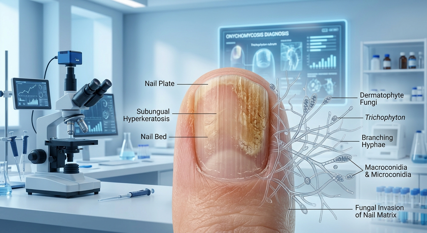
Onychomycosis is a chronic fungal infection of the nail plate, nail bed, or matrix primarily caused by dermatophytes. While often viewed as a cosmetic concern, confirmation of fungal etiology prior to therapy is essential due to non-fungal lookalikes, potential hepatoxicity of systemic antifungals, and severe diabetic limb complications.
Core Clinical Dynamics & Complications
- Etiology: Dermatophytes (predominantly Trichophyton rubrum and Trichophyton mentagrophytes) account for ~90% of toenail onychomycosis cases; non-dermatophyte molds and Candida species account for the remainder.
- Diabetic & High-Risk Complications: Thickened, dystrophic nails in patients with diabetes mellitus, peripheral neuropathy, or peripheral arterial disease can cause subungual ulceration, leading to cellulitis, osteomyelitis, and limb-threatening tissue necrosis.
- Laboratory Confirmation Rule: Always confirm diagnosis via KOH prep, PAS stain, PCR, or fungal culture before initiating systemic oral antifungal therapy.
1. Morphologic Subtypes & Clinical Presentations
| Onychomycosis Subtype | Anatomic & Pathologic Features | Clinical Appearance |
|---|---|---|
| Distal Lateral Subungual (DLSO) | Invasion begins at distal nail bed/hyponychium and moves proximally. Most common pattern. | Subungual hyperkeratosis, yellowish-white discoloration, and onycholysis (separation of nail plate from bed). |
| Endonyx Onychomycosis | Fungal invasion directly into the nail plate interior without involving the underlying nail bed. | Milky-white discoloration of the nail plate without subungual hyperkeratosis or onycholysis. |
| White Superficial (WSO) | Direct invasion of the dorsal surface of the nail plate (often T. mentagrophytes). | Dull, chalky-white patches on the surface of the nail plate that can be easily scraped off with a blade. |
| Total Dystrophic (TDO) | End-stage disease resulting from chronic progression of any subtype. | Complete destruction of the nail unit; thickened, opaque, friable, yellow-brown crumbling nail mass. |
2. Diagnostic Approach & Laboratory Workup
| Diagnostic Modality | Sensitivity / Turnaround | Clinical Utility & Practice Considerations |
|---|---|---|
| Direct Microscopic KOH Exam | Rapid / Moderate Sensitivity (high false-negative rate) | Initial bed-side or clinic test; identifies fungal hyphae/arthrospores but cannot identify specific species or determine viability. |
| Nail Clipping Histopathology (PAS Stain) | Most Sensitive Test / 24–48 hours | Periodic acid–Schiff (PAS) staining of formalin-fixed nail clippings; superior sensitivity compared to KOH and culture for detecting fungal elements. |
| Fungal Culture | High Specificity / 3–4 weeks | Grows pathogen on Sabouraud dextrose agar; essential for identifying specific fungal species and guiding targeted therapy in refractory cases. |
| Polymerase Chain Reaction (PCR) | High Sensitivity & Specificity / 24–48 hours | Rapid identification of fungal DNA; differentiates dermatophytes from molds rapidly without waiting for culture growth. |
| Dermoscopy (Onychoscopy) | Immediate / Non-invasive | Distinguishes fungal etiology (jagged proximal margin with yellow-white spikes) from traumatic onycholysis (smooth linear margin). |
Oral Antifungal Dosing Protocols & Safety Monitoring
Systemic oral therapy yields significantly higher cure rates than topical lacquers for moderate-to-severe toenail involvement.
- Terbinafine (Lamisil®) — First-Line Gold Standard: 250 mg PO daily for 6 weeks (fingernails) or 12 weeks (toenails). Fungicidal against dermatophytes. Baseline LFTs required due to rare risk of hepatotoxicity.
- Itraconazole (Sporanox®) Pulse Dosing: 400 mg PO daily (200 mg BID) for the 1st week of the month, repeated for 2 pulses (fingernails) or 3 pulses (toenails). Drug accumulates in nail matrix for months post-therapy, reducing cost and systemic risk.
- Fluconazole (Diflucan®) Off-Label Alternative: 150 mg to 300 mg PO once weekly for 6 to 12 months.
- Legacy Agent Avoidance: Older agents like Griseofulvin require prolonged administration (up to 18 months), carry low efficacy, and exhibit high relapse rates.
3. Differential Diagnosis & Non-Fungal Mimics
Rule-Out Conditions (Non-Fungal Dystrophies)
- Nail Psoriasis: Features pitting, “oil-drop” discoloration, salmon patches, and symmetric bilateral joint involvement (psoriatic arthritis).
- Traumatic Onycholysis: History of repetitive footwear friction or acute trauma; lacks subungual hyperkeratosis and displays smooth proximal margins on dermoscopy.
- Lichen Planus: Associated with longitudinal ridging, splitting, pterygium formation (dorsal skin adhering to nail bed), and violet cutaneous papules.
- Contact Dermatitis: Periungual inflammation, itching, and scaling caused by acrylic nails or polish ingredients.
High-Yield Exam & Practice Pearls
- Baseline Safety Rule: Always check baseline Liver Function Tests (ALT/AST) prior to initiating oral Terbinafine or Itraconazole.
- The “Treating Without Testing” Trap: ~50% of dystrophic nails are non-fungal in origin (e.g., psoriasis, trauma). Empirical systemic antifungal prescription without diagnostic confirmation is a major clinical pitfall.
- Most Sensitive Single Test: For board examinations, PAS stain of nail clippings is the single most sensitive diagnostic test for confirming onychomycosis.
- Drug Interaction Warning: Itraconazole is a potent CYP3A4 inhibitor and is contraindicated with statins, methadone, and antiarrhythmics. It is also contraindicated in patients with heart failure due to negative inotropic effects.
