
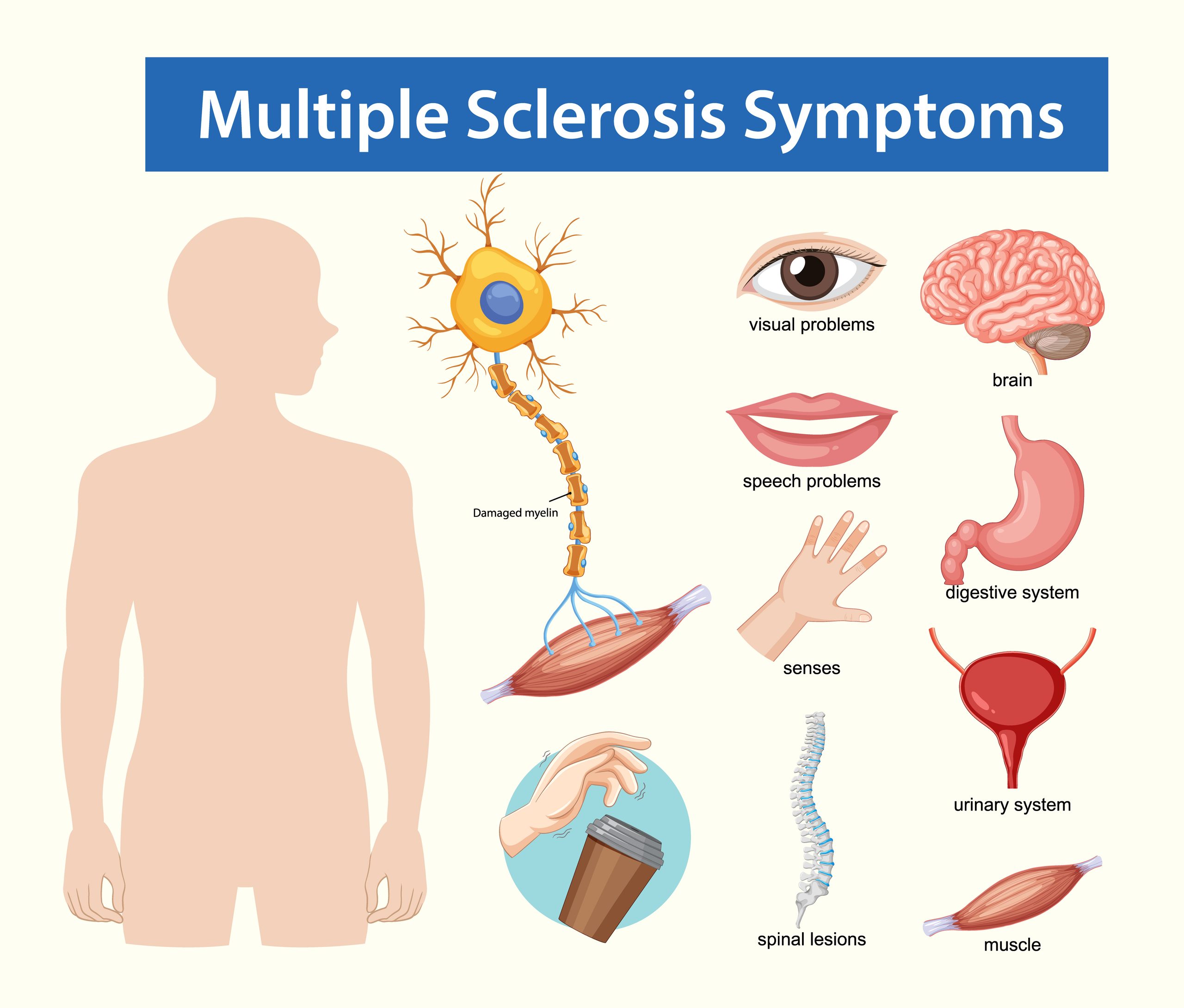
Multiple sclerosis (MS) is a chronic immune-mediated demyelinating disease of the central nervous system (CNS) affecting the brain, spinal cord, and optic nerves. Inflammatory destruction of myelin sheaths impairs impulse conduction and can eventually progress to irreversible axonal and neuronal injury. Hallmark clinical features stem from dissemination in space and time, presenting with highly variable sensory, motor, visual, and autonomic deficits.
Clinical Practice Rule
Establish Dissemination, Exclude Mimics, & Differentiate Pseudo-Relapses: MS diagnosis relies on fulfilling McDonald Criteria for Dissemination in Space (DIS) and Time (DIT) via clinical findings, MRI imaging, and CSF oligoclonal bands. Prior to initiating pulse steroid therapy for acute worsening, clinicians must rule out pseudo-relapses caused by systemic triggers such as urinary tract infections or fever.
1. Epidemiology & Pathophysiology
MS most commonly manifests in young adults aged 20 to 40 years, with women exhibiting a higher incidence of relapsing subtypes. Disease etiology reflects a complex interplay between genetic susceptibility (e.g., HLA-DRB1 alleles) and environmental exposures, including prior Epstein-Barr Virus (EBV) infection, low vitamin D status, higher geographic latitude, and cigarette smoking.
Pathophysiologic Cascade
Normal Myelin Sheath → Autoimmune Leukocyte Infiltration → Acute CNS Demyelination → Conduction Slowing / Block → Repetitive Inflammatory Attacks → Gliosis (Platque Scarring) & Permanent Axonal Loss.
Early inflammatory lesions may undergo partial remyelination and clinical recovery. However, chronic demyelination and persistent inflammation lead to permanent axonal degeneration, brain parenchyma volume loss, and progressive neurological disability.
2. Clinical Subtypes & Phenotypes
| MS Subtype | Disease Course & Pattern | Key Clinical Characteristics |
|---|---|---|
| Relapsing-Remitting (RRMS) | Discrete acute relapses followed by partial or complete recovery (remission). | Most common initial presentation (~85%). Relapses evolve over hours or days, persist for days to weeks, and stabilize or improve. |
| Secondary Progressive (SPMS) | Gradual, progressive decline in function following an initial RRMS disease course. | Accumulation of disability occurs independently of relapses, though occasional superimposed acute attacks may still manifest. |
| Primary Progressive (PPMS) | Continuous worsening of neurological function from disease onset without initial relapses. | Accounts for ~15% of cases. Typically presents at an older median age with progressive myelopathy (e.g., asymmetric spastic paraparesis). |
3. Neuro-Anatomical Symptomatology & Relapse Differentiation
Clinical features mirror the specific anatomical sites of demyelinating lesions across the CNS:
- Optic Neuritis (Optic Nerve): Acute/subacute painful monocular vision loss, decreased visual acuity, central scotoma, and impaired color vision (dyschromatopsia). Ocular motility disorders (e.g., internuclear ophthalmoplegia) reflect brainstem pathway involvement.
- Sensory & Motor (Spinal Cord/Cerebral): Paresthesias, numbness, Lhermitte sign (electric shock-like sensation radiating down the spine on neck flexion), limb weakness, spasticity, hyperreflexia, and gait ataxia.
- Cerebellar & Autonomic: Intention tremor, dysmetria, nystagmus, vertigo, along with neurogenic bladder, bowel dysfunction, sexual dysfunction, and severe central fatigue.
| Clinical Phenomenon | Pathophysiology & Diagnostic Differentiation |
|---|---|
| Uhthoff Phenomenon | Transient worsening of pre-existing neurological deficits triggered by increased core body temperature (e.g., exercise, hot showers, fever). Reflects temperature-dependent conduction block in previously demyelinated axons rather than new tissue damage. Resolves upon cooling. |
| True Acute Relapse | New or acute onset of worsening focal neurological deficits lasting ≥24 hours in the absence of fever, infection, or metabolic derangement. Driven by active immune-mediated CNS inflammation. |
| Pseudo-Relapse | Temporary exacerbation of pre-existing neurological symptoms induced by fever, systemic infection (most commonly Urinary Tract Infections), heat exposure, or physical stress. Does not reflect new CNS lesion formation. |
4. Diagnostic Evaluation & Workup
Diagnosis requires establishing Dissemination in Space (DIS—lesions in ≥2 of 4 typical CNS locations: periventricular, cortical/juxtacortical, infratentorial, spinal cord) and Dissemination in Time (DIT—simultaneous presence of contrast-enhancing and non-enhancing lesions, or a new lesion on follow-up MRI, or CSF-restricted oligoclonal bands).
- Magnetic Resonance Imaging (MRI): Gold standard. Brain and spinal cord T2/FLAIR show characteristic hyperintense demyelinating lesions; T1 with gadolinium identifies active, acute inflammatory breakdown of the blood-brain barrier.
- Cerebrospinal Fluid (CSF) Analysis: Lumbar puncture demonstrates CSF-restricted oligoclonal IgG bands (indicative of intrathecal B-cell antibody synthesis) and an elevated IgG index. Mild mononuclear pleocytosis may be present.
- Visual Evoked Potentials (VEP): Detects subclinical demyelination by measuring prolonged P100 latency along optic pathways.
- Serological Screening: Excludes diagnostic mimics (e.g., Aquaporin-4 IgG for NMOSD, MOG-IgG for MOGAD, B12 deficiency, neurosyphilis, and systemic autoimmune vasculitis).
5. Therapeutic Management & Disease-Modifying Therapies (DMTs)
A. Acute Relapse Management
- High-Dose Corticosteroids: Intravenous methylprednisolone (1,000 mg daily for 3 to 5 days) accelerates functional recovery by reducing acute focal tissue edema and capillary permeability. (Note: Steroids do not alter long-term disability).
- Plasma Exchange (PLEX): Indicated as a second-line rescue intervention for severe, fulminant demyelinating relapses refractory to high-dose IV corticosteroid pulse therapy.
B. Disease-Modifying Therapies (DMTs)
| Therapy / Class | Mechanism of Action | Key Clinical Safety & Monitoring Considerations |
|---|---|---|
| Injectables (Interferon beta, Glatiramer acetate) |
Injectable immunomodulators; shifts cytokine balance and alters T-cell reactivity against myelin basic protein. | Flu-like syndrome, injection-site necrosis, hepatic enzyme elevations, and cytopenias. Lower overall efficacy compared to newer oral/infusion agents. |
| Oral Immunomodulators (Dimethyl fumarate, Teriflunomide) |
Nuclear factor-like 2 (Nrf2) pathway activation / Pyrimidine synthesis inhibition reducing lymphocyte proliferation. | GI intolerance, flushing, lymphopenia. Teriflunomide carries boxed warnings for severe hepatotoxicity and teratogenicity (requires accelerated elimination protocol if pregnancy is desired). |
| S1P Modulators (Fingolimod, Siponimod) |
Sphingosine-1-phosphate receptor modulators; sequesters lymphocytes inside peripheral lymph nodes. | First-dose sinus bradycardia/AV block (requires 6-hour cardiac monitoring), macular edema, varicella zoster reactivation, and risk of severe rebound disease upon abrupt cessation. |
| Integrin Blockers (Natalizumab) |
Monoclonal antibody targeting α4-integrin; blocks leukocyte adhesion and extravasation across the blood-brain barrier. | High risk of Progressive Multifocal Leukoencephalopathy (PML) caused by JC virus reactivation. Requires strict risk stratification via anti-JCV antibody index testing. |
| Anti-CD20 Agents (Ocrelizumab, Ofatumumab) |
Monoclonal antibodies binding CD20; leads to selective depletion of circulating CD20+ B cells. | Approved for both RRMS and PPMS. Infusion/injection-related reactions, increased susceptibility to upper respiratory and herpesvirus infections, and hypogammaglobulinemia. |
| Immune Reconstitution (Alemtuzumab, Cladribine) |
Anti-CD52 monoclonal antibody / purine nucleoside analog inducing rapid, profound depletion of T and B lymphocytes. | High incidence of secondary autoimmune disorders (e.g., Immune Thrombocytopenia, thyroid disease, anti-GBM disease). Requires prolonged laboratory monitoring for years post-dosing. |
Clinical Practice Pearls
- Always Rule Out Infection in Suspected Relapses: Perform a urinalysis and basic infection screening before diagnosing an acute inflammatory relapse to avoid inappropriate steroid pulse therapy for a pseudo-relapse.
- PML Risk Stratification: In patients receiving Natalizumab, monitor anti-JCV antibody titers every 6 months. A positive status with high index values warrants reassessment of the benefit-risk ratio or transition to an alternative high-efficacy DMT.
- PPMS Therapeutic Selection: Ocrelizumab remains the primary FDA-approved disease-modifying agent shown to delay disability progression in patients with Primary Progressive MS.
