
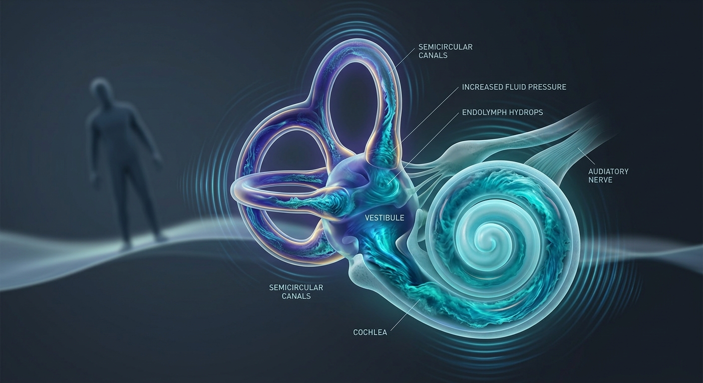
Ménière’s disease and Ménière’s syndrome share an identical symptom complex characterized by inner-ear dysfunction. The critical diagnostic boundary rests entirely on etiology: Ménière’s disease is idiopathic, whereas Ménière’s syndrome stems from an identifiable underlying pathology.
Etiology & Core Pathophysiologic Mechanism
- Ménière’s Disease vs. Syndrome:
- Ménière’s Disease: Idiopathic endolymphatic hydrops without a recognizable secondary cause. Peak incidence occurs between 40–60 years of age with a slight female predominance.
- Ménière’s Syndrome: Endolymphatic hydrops secondary to a known systemic or localized disorder (e.g., systemic lupus erythematosus [SLE], rheumatoid arthritis [RA], otosclerosis, or post-traumatic/viral labyrinthitis).
- Core Mechanism (Endolymphatic Hydrops):
↑ Endolymphatic Fluid Volume/Pressure → Membrane Distortion & Rupture → Dual Dysfunction: Cochlear System (Fluctuating Hearing Loss & Tinnitus) + Vestibular System (Vertigo & Imbalance) - Common Flare Triggers: High dietary sodium, caffeine, alcohol, elevated physical/emotional stress, and menstrual cycle variations.
1. The Classic Diagnostic Tetrad
| Tetrad Feature | Clinical Presentation & Progression | Key Diagnostic Pearls |
|---|---|---|
| 1. Episodic Vertigo | Recurrent room-spinning vertigo lasting minutes to hours (typically 20 min to 24 hours). Accompanied by severe nausea, profuse vomiting, and post-episode exhaustion. | Must be distinguished from brief position-induced episodes (<1 min in BPPV) or continuous multi-day vertigo (vestibular neuritis). |
| 2. Fluctuating Hearing Loss | Progressive Sensorineural Hearing Loss (SNHL) that heralds or accompanies acute vertigo flares. Early loss affects low-to-moderate frequencies; advances to high frequencies over time. | Unilateral in early disease; audiometry confirms SNHL. Reversible initially, but permanent loss accrues over years. |
| 3. Tinnitus | Low-pitched ringing, roaring, or buzzing tone that characteristically fluctuates in intensity prior to or during an acute attack. | Roaring character distinguishes it from high-pitched tonal tinnitus common in presbycusis or noise exposure. |
| 4. Aural Fullness | Deep, uncomfortable sensation of pressure, fullness, or a “plugged” ear on the affected side prior to or during exacerbations. | Reflects increased hydraulic pressure within the membranous labyrinth. |
2. Bedside & Diagnostic Examination Strategies
| Diagnostic Test | Expected Clinical Finding | Diagnostic Significance & Interpretation |
|---|---|---|
| Weber Tuning Fork Test | Sound lateralizes to the unaffected (better) ear. | Confirms sensorineural hearing loss in the affected ear (sound travels to the functional inner ear). |
| Rinne Tuning Fork Test | Air Conduction > Bone Conduction (AC > BC) bilaterally. | Normal/Positive result; excludes conductive middle-ear pathology. |
| Fukuda Marching Step Test | Progressive rotational drift toward the affected (faulty) ear. | Identifies unilateral vestibular tone deficit as the patient marches in place with eyes closed. |
| Romberg Test | Positive (increased unsteadiness/sway with eyes closed). | Demonstrates proprioceptive and vestibular imbalance. |
| Pneumatic Otoscopy | Pressure changes elicit vertigo or nystagmus (Hennebert’s sign). | Suggests abnormal mechanical coupling between the middle and inner ear. |
| Dix-Hallpike Maneuver | Typically negative; may be weakly positive. | A strongly positive result with classic torsional nystagmus indicates coexisting BPPV. |
| Neuroimaging (CT/MRI) | Unremarkable for structural defects. | Not routinely required unless needed to rule out central pathology or vestibular schwannoma (acoustic neuroma). |
3. Phase-Specific Management & Treatment Protocols
| Phase / Goal | Therapeutic Class & Agents | Clinical Mechanics & Safety Warnings |
|---|---|---|
| Acute Attack Control Rescue Therapy |
• Vestibular Suppressants: Meclizine (Antihistamine) or Benzodiazepines (Diazepam). • Antiemetics: Ondansetron or Promethazine. • Corticosteroids: Short-term oral Prednisone taper. |
Controls disabling vertigo, nausea, and anxiety during acute attacks. Corticosteroids reduce inner-ear endolymphatic inflammation and preserve hearing. Does not prevent future attacks. |
| Attack Prevention Maintenance |
• Diuretics: Thiazides (e.g., Hydrochlorothiazide/Triamterene). • Lifestyle Modification: Low-salt diet (<2,000 mg/day), caffeine & alcohol restriction, stress reduction. |
Lowers endolymphatic volume and hydraulic pressure. Thiazides are for prophylaxis only; they have no role in terminating an acute attack. |
| Refractory / Invasive ENT Options Specialist Level |
• Conservative Procedures: Endolymphatic sac decompression. • Ablative Therapies: Intratympanic Gentamicin (chemical labyrinthectomy), vestibular neurectomy, or surgical labyrinthectomy. |
Reserved for intractable, disabling vertigo failing conservative measures. Warning: Ablative procedures carry a high risk of permanent sensorineural hearing loss. |
| Secondary Disease Control | Targeted therapy for underlying disorder (e.g., immunosuppressants for SLE/RA). | Essential in Ménière’s syndrome to halt ongoing secondary labyrinthine damage. |
4. High-Yield Clinical Comparisons
| Clinical Feature | Ménière’s Disease | Benign Paroxysmal Positional Vertigo (BPPV) |
|---|---|---|
| Vertigo Duration | Episodic, lasting minutes to hours (20 min – 24 hr). | Brief paroxysmal attacks, lasting <1 minute. |
| Auditory Symptoms | PRESENT ⭐ (Hearing loss, tinnitus, aural fullness). | ABSENT. |
| Dix-Hallpike Test | Usually negative (unless concurrent BPPV). | Classically Diagnostic (rotational nystagmus with latency). |
| Primary Triggers | Sodium intake, stress, or spontaneous pressure shifts. | Specific head-position changes (e.g., rolling over in bed, looking up). |
Primary Clinical Separator
Vertigo paired with auditory symptoms (hearing loss, tinnitus, or aural fullness) points directly to Ménière’s Disease/Syndrome rather than BPPV or vestibular neuritis.
High-Yield Exam & Practice Pearls
- Disease vs. Syndrome: Disease = Idiopathic endolymphatic hydrops; Syndrome = Secondary endolymphatic hydrops (e.g., SLE, RA).
- The Classic Tetrad: Episodic vertigo + fluctuating SNHL + tinnitus + aural fullness.
- Tuning Fork Pattern: Sensorineural loss → Weber lateralizes to the UNAFFECTED (good) ear; Rinne is normal bilaterally (AC > BC).
- Gait Directionality: Fukuda step test shows rotational drift toward the AFFECTED (faulty) ear.
- Diuretic Utility: Thiazide diuretics are strictly for prevention of attacks; they are useless for acute symptom termination.
- Rescue Therapy: Meclizine, antiemetics, benzodiazepines, and short courses of steroids manage acute exacerbations.
