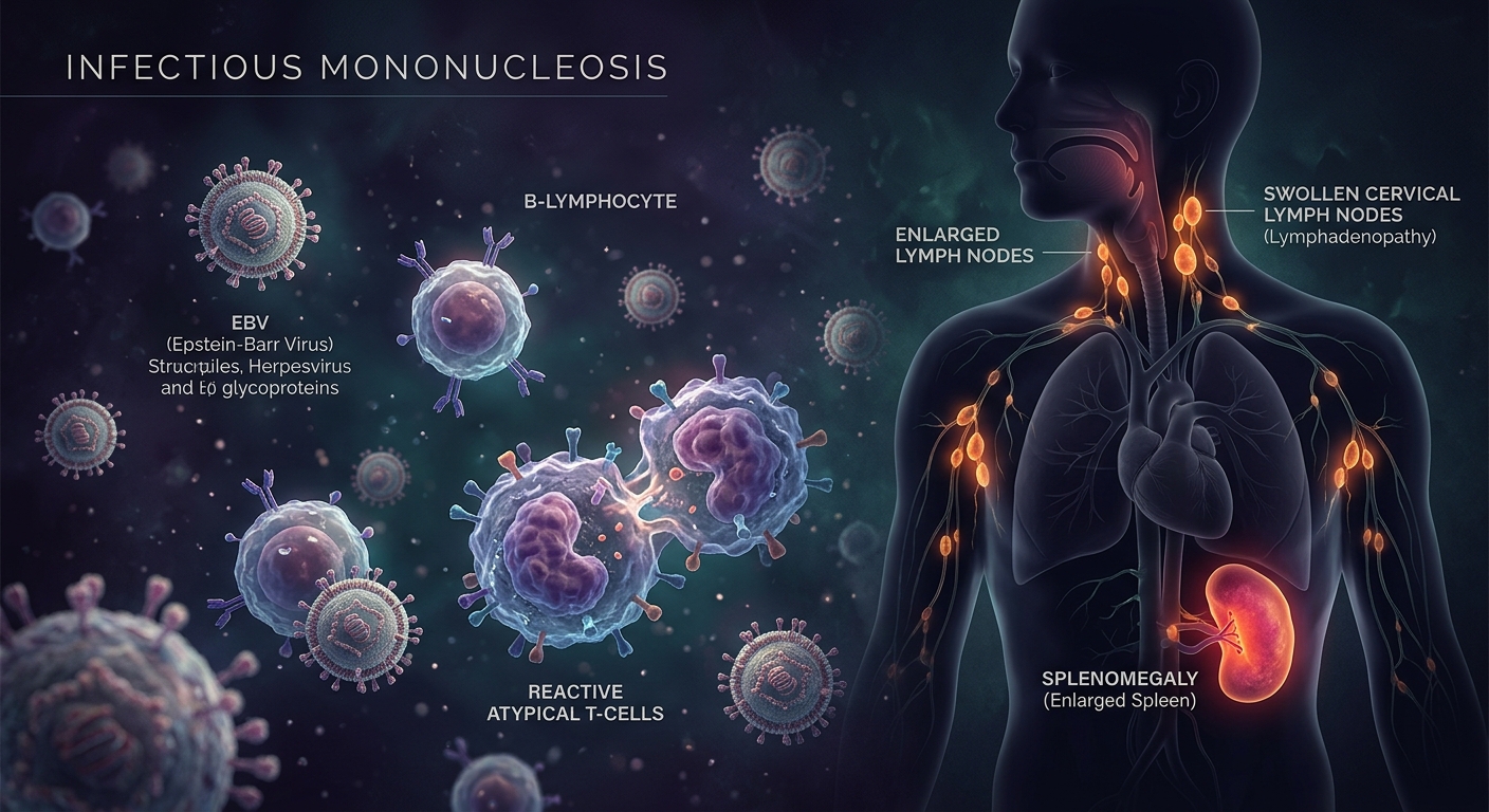
Infectious Mononucleosis (IM) is an acute systemic viral syndrome predominantly caused by the Epstein-Barr virus (EBV), a double-stranded DNA herpesvirus. Commonly known as the “kissing disease,” EBV is transmitted via oropharyngeal secretions (saliva) and preferentially infects B lymphocytes. While most cases occur in patients under 35 years old, distinguishing IM from bacterial pharyngitis is critical to prevent inappropriate antibiotic use, avoid severe cutaneous drug reactions, and minimize the risk of life-threatening splenic rupture.
Clinical Course & Key Examination Findings
- Predictable Timeline:
- Prodrome (3–5 Days): Headaches, severe malaise, myalgias, and anorexia.
- Acute Phase (5–15 Days): Classic triad of Fever + Exudative Pharyngitis + Lymphadenopathy paired with debilitating fatigue.
- Convalescence (4–6+ Weeks): Slow resolution; profound fatigue may linger for months.
- Prominent Physical Exam Signs:
- Posterior Cervical Adenopathy: Prominence in posterior cervical chains or generalized lymphadenopathy strongly favors EBV over GAS.
- Splenomegaly (>50% of cases): Tender, enlarged spleen presenting maximum rupture risk during weeks 2–3.
- Hepatomegaly (~10% of cases): Tender RUQ with transient jaundice or mild transaminitis.
- Mucocutaneous Signs: Soft-palate petechiae, periorbital edema, and transient maculopapular exanthems.
1. Differential Diagnosis: EBV Mono vs. GAS Pharyngitis
| Clinical Feature |
Infectious Mononucleosis (EBV) |
GAS Pharyngitis (S. pyogenes) |
| Primary Etiology |
Epstein-Barr Virus (DNA Herpesvirus) |
Streptococcus pyogenes (GABHS) |
| Fatigue Severity |
Prominent / Severe (weeks to months) |
Moderate / Mild |
| Lymphadenopathy Location |
Posterior Cervical & Generalized |
Anterior Cervical primarily |
| Splenomegaly / Hepatomegaly |
Common (>50% Spleen / ~10% Liver) |
Absent |
| Aminopenicillin Response |
Classic Maculopapular Rash |
Therapeutic clearance (No rash) |
| Duration of Illness |
Protracted (4–6 weeks) |
Short (resolves rapidly with antibiotics) |
2. Diagnostic Testing & Laboratory Workup
| Diagnostic Test |
Key Findings & Mechanics |
Diagnostic Pitfalls & Notes |
| Heterophile Antibody (Monospot) |
Detects heterophile antibodies produced by EBV-stimulated B cells (Sensitivity ~85%, Specificity ~100%). |
Early False Negatives: Only ~60% positive in Week 1. Up to 50% false-negative in children <4 years. |
| CBC & Peripheral Smear |
Leukopenia followed by absolute lymphocytosis; atypical lymphocytes (reactive CD8+ T cells / Downey cells). |
Mild thrombocytopenia observed in ~50% of uncomplicated cases. |
| Hepatic Transaminases |
Elevated AST & ALT (2–3× upper limit of normal) in ~85% of patients. |
Transaminitis typically peaks between weeks 2 and 3 of infection. |
The Amoxicillin / Ampicillin Rash Pitfall
Administering Amoxicillin or Ampicillin during active EBV infection triggers a characteristic generalized maculopapular rash in up to 90% of cases. This reaction is mediated by transient EBV-induced immune dysregulation and does NOT indicate a true IgE-mediated penicillin allergy.
3. Therapeutic Management & Activity Restrictions
| Intervention / Category |
Clinical Guidance |
Key Exceptions & Restrictions |
| First-Line Therapy |
Supportive Care: Rest, hydration, NSAIDs / Acetaminophen for fever and sore throat. |
Gradual return to daily activity based on symptom resolution. |
| Antiviral Therapy |
NOT ROUTINELY INDICATED |
Acyclovir decreases viral shedding but provides no meaningful clinical benefit. |
| Corticosteroid Use |
Indicated ONLY for Airway Compromise (e.g., Prednisone 40–60 mg/day PO for 3 days). |
Reserved for impending upper airway obstruction due to massive tonsillar hypertrophy. |
| Activity Restrictions |
Strict avoidance of contact sports, heavy lifting, and collision activities. |
Minimum 1 Month (28 Days) Restriction due to highest splenic rupture risk in weeks 2–3. |
High-Yield Exam & Practice Pearls
- Target Cell Mechanism: EBV infects B lymphocytes via the CD21 receptor.
- Classic Diagnostic Triad: Fever + Exudative Pharyngitis + Cervical Lymphadenopathy (especially posterior chain).
- Systemic Footprint: Distinct from localized strep; features splenomegaly (>50%) and elevated AST/ALT (~85%).
- Monospot Timing: High false-negative rate during the first week. Early negative tests require repeat testing if clinical suspicion persists.
- Smear Findings: Reactive “atypical lymphocytes” (Downey cells) on blood smear represent CD8+ cytotoxic T cells attacking infected B cells.
- Steroid Mandate: Systemic steroids are contraindicated for routine mono; reserve exclusively for severe upper airway obstruction or severe autoimmune cytopenias.
- Drug Reaction: Amoxicillin + EBV = Maculopapular Rash (viral-mediated immune reaction, not a true IgE allergy).
- Rupture Window: Peak incidence of splenic rupture is during weeks 2–3; enforce a strict minimum 1-month sports restriction.