
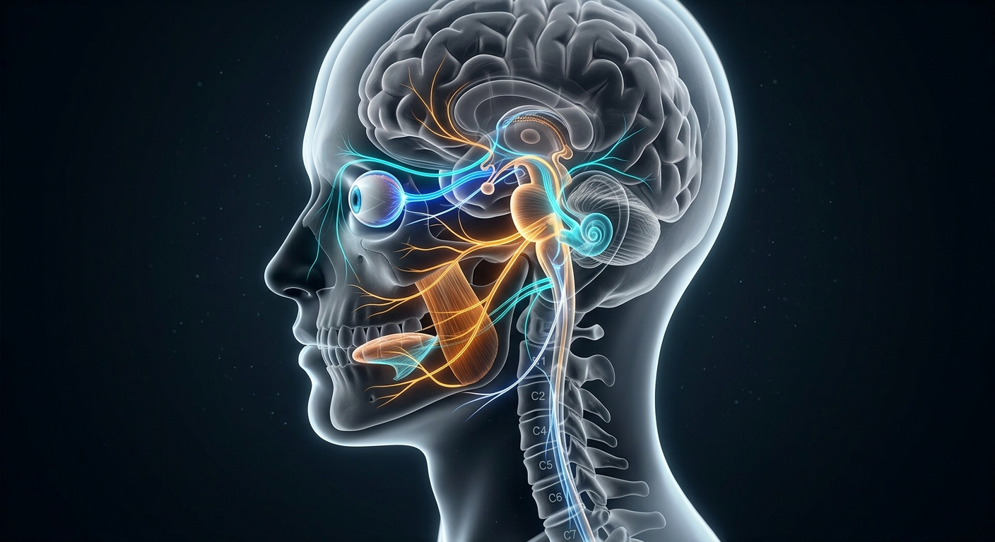
The twelve pairs of cranial nerves are critical structures of the peripheral nervous system, mediating sensory, motor, and parasympathetic control across the head, neck, and visceral organs. Evaluating cranial nerve function provides localized diagnostic insight into brainstem lesions, vascular events, or peripheral nerve pathologies, making standard cranial nerve assessment a foundational clinical competency.
Standard Clinical Mnemonics
- Nerve Names Order: “On Old Olympus Towering Tops, A Finn And German Viewed Some Hops”
- Alternative Names Order: “Oh Oh Oh To Touch And Feel A Great Vein, Ah Heaven!”
- Functional Classification (Sensory/Motor/Both): “Some Say Marry Money But My Brother Says Big Brains Matter More” (S = Sensory, M = Motor, B = Both)
Master Reference Table: The 12 Cranial Nerves
| Number & Name | Type | Primary Function | Assessment Method | Clinical Findings & Deficits |
|---|---|---|---|---|
| CN I: Olfactory | Sensory | Olfaction (Smell) | Test each nostril individually with non-irritating scents. | Anosmia (loss of smell) |
| CN II: Optic | Sensory | Vision; afferent limb of pupillary reflex | Snellen chart, visual fields by confrontation, pupillary light response. | Visual field defects, loss of acuity, afferent pupillary defect |
| CN III: Oculomotor | Motor | Most extraocular movements, eyelid elevation, pupillary constriction | Inspect eyelid symmetry, extraocular movements, pupillary light response. | Ptosis, “down and out” resting eye position, dilated non-reactive pupil |
| CN IV: Trochlear | Motor | Superior oblique muscle (inward & downward gaze) | Evaluate downward and inward ocular tracking. | Vertical diplopia (worse reading or descending stairs) |
| CN V: Trigeminal | Both | Facial sensation (V1, V2, V3); muscles of mastication | Facial light touch/pain, jaw clench, corneal reflex afferent. | Facial anesthesia, masseter/temporalis weakness, absent corneal reflex |
| CN VI: Abducens | Motor | Lateral rectus muscle (ocular abduction) | Assess horizontal lateral gaze. | Inability to abduct eye, convergent squint, horizontal diplopia |
| CN VII: Facial | Both | Facial expression, anterior 2/3 taste, lacrimation, salivation | Smile, raise eyebrows, puff cheeks, close eyes against resistance. | Facial asymmetry, loss of taste, Bell’s palsy (lower motor neuron lesion) |
| CN VIII: Vestibulocochlear | Sensory | Auditory perception & vestibular equilibrium | Whisper voice test, Rinne and Weber tuning fork tests, Romberg test. | Sensorineural hearing loss, tinnitus, vertigo, gait ataxia |
| CN IX: Glossopharyngeal | Both | Posterior 1/3 taste, swallowing, carotid baroreceptors | Assess gag reflex, palate elevation, deglutition. | Dysphagia, impaired gag reflex, loss of posterior tongue taste |
| CN X: Vagus | Both | Parasympathetic thoracic/abdominal visceral control, phonation, swallowing | Voice quality (hoarseness), symmetrical palate elevation (“ah”), gag reflex. | Hoarseness, uvula deviation (away from lesion site), dysphagia, visceral autonomic impairment |
| CN XI: Spinal Accessory | Motor | Trapezius & sternocleidomastoid motor innervation | Shrug shoulders and rotate head against manual resistance. | Asymmetrical shoulder shrug, weakness rotating head to contralateral side |
| CN XII: Hypoglossal | Motor | Intrinsic and extrinsic tongue musculature | Inspect tongue protrusion and rapid lateral movements. | Tongue deviation toward affected (weak) side, dysarthria |
Anatomical & Functional Breakdown
Trigeminal Nerve (CN V) Anatomical Divisions
| Division | Function Type | Anatomical Distribution |
|---|---|---|
| Ophthalmic (V1) | Sensory | Forehead, cornea, upper eyelid, bridge of nose. |
| Maxillary (V2) | Sensory | Cheeks, upper teeth, nasal cavity, lower eyelid. |
| Mandibular (V3) | Sensory & Motor | Sensory: Lower face, chin, lower teeth. Motor: Muscles of mastication (masseter, temporalis, pterygoids). |
Integrated Reflex Loops & Ocular Systems
The Corneal Reflex Arc
A protective reflex pathway requiring dual-nerve coordination:
- Afferent (Sensory) Limb: Mediated by the Ophthalmic division (V1) of CN V (Trigeminal), detecting corneal contact.
- Efferent (Motor) Limb: Mediated by CN VII (Facial), stimulating bilateral orbicularis oculi contraction to produce rapid blinking.
Extraocular Eye Movement (EOM) Motor System
| Cranial Nerve | Innervated Musculature | Primary Direction of Gaze |
|---|---|---|
| CN III (Oculomotor) | Superior, Inferior, & Medial Recti; Inferior Oblique | Upward, downward, and inward gaze |
| CN IV (Trochlear) | Superior Oblique | Downward and inward gaze |
| CN VI (Abducens) | Lateral Rectus | Outward (lateral abduction) gaze |
Clinical Practice Pearls
- Distinguish Upper vs. Lower Motor Neuron CN VII Lesions: UMN lesions (e.g., stroke) spare the forehead due to bilateral cortical representation; LMN lesions (e.g., Bell’s palsy) cause total ipsilateral hemifacial paralysis.
- Remember the Uvula Deviation Rule: In CN X palsies, the soft palate fails to elevate on the affected side, causing the uvula to deviate away from the lesion toward the healthy side.
- Tongue Points to the Lesion: In CN XII motor weakness, the protruded tongue deviates toward the weak (affected) side due to unopposed action of the contralateral genioglossus muscle.
