
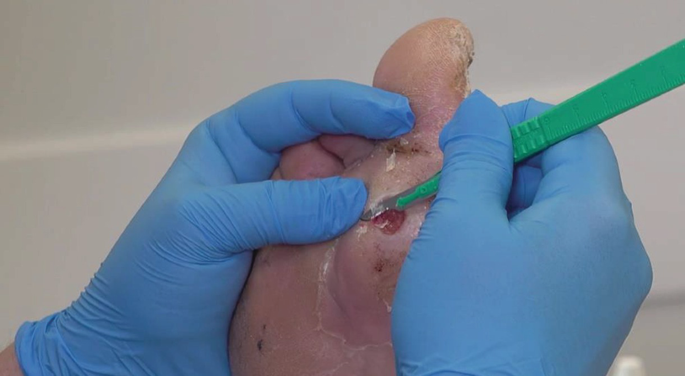
Conservative Sharp Wound Debridement (CSWD) is a specialized bedside procedure involving the deliberate, controlled excision of non-viable, necrotic tissue, slough, or bioburden using sharp instruments strictly above the level of viable tissue. While CSWD rapidly transforms stalled wound environments to accelerate healing, it carries explicit clinical risks and demands specialized competence, formal mentorship, and institutional authorization.
CSWD Quick Reference Guide
| Primary Objective | Excision of devitalized tissue, pus, slough, or foreign material to optimize the wound bed. |
| Common Instrumentation | Scalpel, tissue scissors, dermal curette, dissection forceps. |
| Comparative Advantage | Fastest debridement method; highly effective for heavy exudate and high microbial burden. |
| Core Safety Rule | When in doubt, leave it alone. Never debride tissue that cannot be clearly visualized. |
Selecting the Appropriate Debridement Modality
Choosing CSWD over alternative methods requires balancing clinical urgency against patient factors. Wounds Canada ranks modalities across key clinical performance indicators:
| Debridement Method | Speed | Tissue Selectivity | Pain Level | Exudate Management | Infection Control | Cost Efficiency |
|---|---|---|---|---|---|---|
| Surgical / Sharp (CSWD) | 1 (Fastest) | 3 | 5 (Highest) | 1 (Best) | 1 (Best) | 5 |
| Biological (Biosurgical) | 2 | 2 | 3 | 5 | 2 | 3 |
| Enzymatic | 3 | 1 (Most Selective) | 2 | 4 | 4 | 2 |
| Mechanical | 4 | 5 | 4 | 2 | 3 | 4 |
| Autolytic | 5 (Slowest) | 4 | 1 (Lowest) | 3 | 5 | 1 (Lowest) |
Wounds Canada comparative scale: 1 = Most Desirable, 5 = Least Desirable.
Clear Clinical Contraindications
Absolute & Relative Contraindications
Do NOT proceed with Conservative Sharp Wound Debridement under the following clinical conditions:
| Condition | Clinical Rationale |
|---|---|
| Poorly Visualized Tissue | Structures beneath opaque slough, deep sinus tracts, or poorly lit areas pose severe risk of inadvertent vascular or structural injury. |
| Dry Eschar with Ischemia | Stable, dry eschar on ischemic extremities (minimal perfusion) serves as a natural barrier. Sharp removal risks unmasking non-healing ulcers and introducing infection. |
| Radiated Wound Beds | Irradiated tissue exhibits altered microvascularity and compromised tissue regeneration, raising the risk of non-healing surgical defects. |
| Vasculitic Wounds | Active vasculitis causes severe small-vessel inflammation; sharp intervention can trigger intense pathergy and wound expansion. |
| Clinician Uncertainty | Lack of clear structural identification, inadequate skill/authorization, or missing emergency supplies requires immediate pause. |
Structured 7-Step CSWD Clinical Workflow
| Step | Clinical Protocol | Key Safety Actions |
|---|---|---|
| 1. Assess Safety | Verify clinical scope, institutional policy support, clinician competence, and environment. | Confirm proper lighting, ergonomics, assistant availability, and sharps disposal containers. |
| 2. Patient & Wound Assessment | Perform a thorough wound evaluation (etiology, tissue types, perfusion) and baseline pain assessment. | Utilize validated assessment tools for pain and wound parameters before proceeding. |
| 3. Set the Stage | Discuss procedure rationale, anticipated benefits, risks, and alternatives with the patient. | Obtain and document verbal or written informed consent per institutional policy. |
| 4. Assemble Resources | Gather sterile dressing tray, instruments (scalpel, curette, scissors), protective pads, and PPE. | Bleeding Preparedness: Have hemostatic agents, pressure dressings, and silver nitrate ready before making the first incision. |
| 5. Perform CSWD | Excise non-viable tissue conservatively using sharp instruments strictly above viable planes. | Work within clearly identified boundaries. Stop immediately if viable, vascularized tissue is encountered or if unmanageable pain occurs. |
| 6. Post-Procedure Care | Apply targeted secondary dressing, manage residual pain, dispose of sharps safely, and reassess patient status. | Re-measure the wound: Removing tissue alters wound dimensions—update baseline length, width, and depth in charts. |
| 7. Evaluate Outcomes | Determine if debridement achieved the desired clinical goals (e.g., bioburden reduction, exudate control). | If progress stalls or goals are unmet, re-evaluate etiology and transition to alternative debridement strategies. |
Managing Bleeding Risk & Complications
Bleeding during conservative sharp debridement indicates penetration into viable, vascularized tissue—which falls outside the scope of non-surgical CSWD and must be treated as an incident:
Bleeding Response Protocol
- Immediate Hemostasis: Apply direct pressure with sterile gauze for a minimum of 5 to 10 minutes.
- Topical Hemostats: Utilize calcium alginate dressings, silver nitrate applicators, or topical hemostatic agents if light capillary oozing persists.
- Escalation: If arterial or brisk venous bleeding occurs, maintain continuous firm pressure, notify the attending physician or surgical team immediately, and initiate emergency protocols.
- Incident Reporting: Document the event, volume of blood loss, interventions performed, and submit an internal safety incident report per facility standards.
Post-Procedure Documentation Requirements
Comprehensive charting protects both patient safety and clinical accountability. Document the following items immediately post-procedure:
- Informed consent verification and pre-procedure pain score.
- Pre-debridement tissue characteristics (percentage slough, eschar, exudate volume).
- Specific instruments used and anatomical boundaries respected.
- Type and approximate volume/amount of non-viable tissue excised.
- Intra-procedure pain tolerance and interventions (e.g., topical analgesics applied).
- Complications encountered (e.g., capillary bleeding) and hemostatic measures used.
- Post-debridement tissue appearance and updated wound measurements.
- Primary and secondary dressings applied, along with follow-up plan.
Clinical Practice Pearls
- Respect Blood Supply: Never perform CSWD on dry, gangrenous digits or ischemic wounds without verified vascular clearance.
- Manage Pain Preemptively: Administer ordered systemic or topical analgesics early enough to allow full therapeutic onset prior to debridement.
- Re-Measure Every Time: Always record new wound dimensions post-debridement, as tissue removal routinely alters perimeter and depth measurements.
