
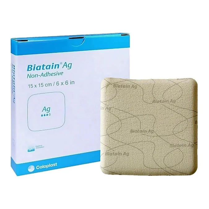
Biatain® Ag Non-Adhesive is an advanced antimicrobial polyurethane foam dressing engineered for moderate-to-highly exuding wounds facing local infection, suspected biofilm, or elevated risk of contamination. Combining dynamic exudate management with continuous silver release, this non-adhesive format is designed to protect fragile periwound skin while eliminating pooling in the wound bed.
Key Technical Specifications & Overview
| Dressing Structure | Silver-containing polyurethane foam with semi-permeable film backing |
| Core Technology | 3DFit® Technology (conforms vertically to wound beds up to 2 cm depth) |
| Antimicrobial Activity | Sustained silver ion release for up to 7 days; effective against mature biofilm |
| Fixation Required | Non-adhesive (requires retention tape, bandage, or compression) |
Mechanism of Action: How Biatain Ag Works
Biatain Ag Non-Adhesive integrates vertical exudate absorption with controlled antimicrobial delivery:
- Vertical Exudate Absorption: The absorbent foam lock-in mechanism pulls exudate straight up, keeping fluid away from vulnerable wound edges to prevent maceration.
- 3DFit® Conformability: Upon contact with exudate, the foam expands vertically to conform directly to the wound bed (up to ~2 cm depth), minimizing dead space where bacteria pool.
- Sustained Ionic Silver Release: Wound fluid activates silver ions within the foam matrix, providing continuous antimicrobial defense for up to 7 days.
- Biofilm Disruption: In-vitro testing demonstrates active disruption of mature bacterial biofilms and prevention of new biofilm development.
- Protective Barrier: The semi-permeable outer film defends against external pathogens while facilitating optimal moisture vapor exchange.
Clinical Indications
Biatain Ag Non-Adhesive is indicated for exuding superficial to moderately deep wounds requiring antimicrobial therapy:
- Localized Infection: Indicated when presenting with localized signs such as unexpected pain, malodor, altered exudate, delayed healing, friable tissue, or localized erythema.
- Suspected Biofilm: Recommended for chronic, stalled wounds that fail to progress despite standard care or show recurring infection cycles.
- High-Risk Wounds: Applied prophylactically in selected non-healing wounds vulnerable to contamination.
- Diabetic Foot Ulcers (DFUs): The non-adhesive structure prevents skin tearing on friable periwound tissue (requires offloading and perfusion assessment).
- Venous Leg Ulcers (VLUs): Safe and effective for use underneath multi-layer compression therapy systems following vascular clearance.
Precautions & Contraindications
Contraindications
- Silver Hypersensitivity: Do not use in patients with known allergy or sensitivity to silver or polyurethane foam components.
- Oxidizing Cleansers: Avoid combining directly with hydrogen peroxide or hypochlorite solutions (use sterile saline or water instead).
Precautions & Special Procedures
| Scenario | Clinical Recommendation |
|---|---|
| Therapeutic Radiation & X-Ray | Remove the dressing prior to treatment/imaging and replace afterward as per clinical protocol. |
| Electrical Procedures & Diathermy | Avoid direct contact between silver dressing and electrodes or conductive gels. |
| MRI Procedures | May remain in place during MRI scans when permitted by manufacturer and institutional policies. |
| Pregnancy, Lactation & Pediatrics | Use cautiously based on individualized clinical assessment and manufacturer guidelines. |
| Tissue Discoloration | Transient, benign skin/wound bed discoloration may occur due to silver content. |
Step-by-Step Application Protocol
| Phase | Clinical Procedure | Clinical Pearls |
|---|---|---|
| 1. Assessment | Evaluate wound size, depth (up to 2 cm), exudate volume, periwound condition, pain, and signs of spreading infection. | If infection extends systemically, systemic antibiotics must accompany topical care. |
| 2. Cleansing | Irrigate wound with sterile normal saline or sterile water. Thoroughly dry the periwound area. Apply barrier film if needed. | Keeping periwound skin dry prevents moisture damage and ensures secure fixation tape placement. |
| 3. Sizing | Select a size providing at least a 2 cm overlap beyond wound margins (1 cm overlap for very small wounds). | Adequate overlap allows room for foam expansion and prevents peripheral leakage. |
| 4. Placement & Fixation | Place the plain, non-printed side directly against the wound bed. Secure with tape, conforming wrap, or compression. | Note Lightly moisten foam with sterile saline prior to application only if exudate levels are very low. |
| 5. Removal | Gently lift corners and remove carefully. If the dressing adheres, irrigate with sterile saline to soak free. | Never forcibly remove adherent foam. Repeated sticking indicates low exudate—reassess if foam is still appropriate. |
Wear Time & Expected Outcomes
- Maximum Wear Time: Up to 7 days depending on exudate levels and infection status. Change immediately if fluid reaches the foam edge or leakage occurs.
- Infection Resolution: Signs of localized infection should show noticeable improvement with target resolution within 2 weeks.
- Re-evaluation Rule: If no improvement occurs within 14 days, reassess vascular status, need for surgical debridement, offloading, or systemic infection escalation.
Practice Pearls for Clinicians
- Orientation Check: Always place the non-printed surface toward the patient’s wound bed.
- Fixation Selection: Pair with retention bandages or silicon tape when managing fragile or paper-thin skin.
- Silver Stewardship: Re-evaluate silver therapy regularly; avoid indefinite routine use once bioburden is controlled.
