
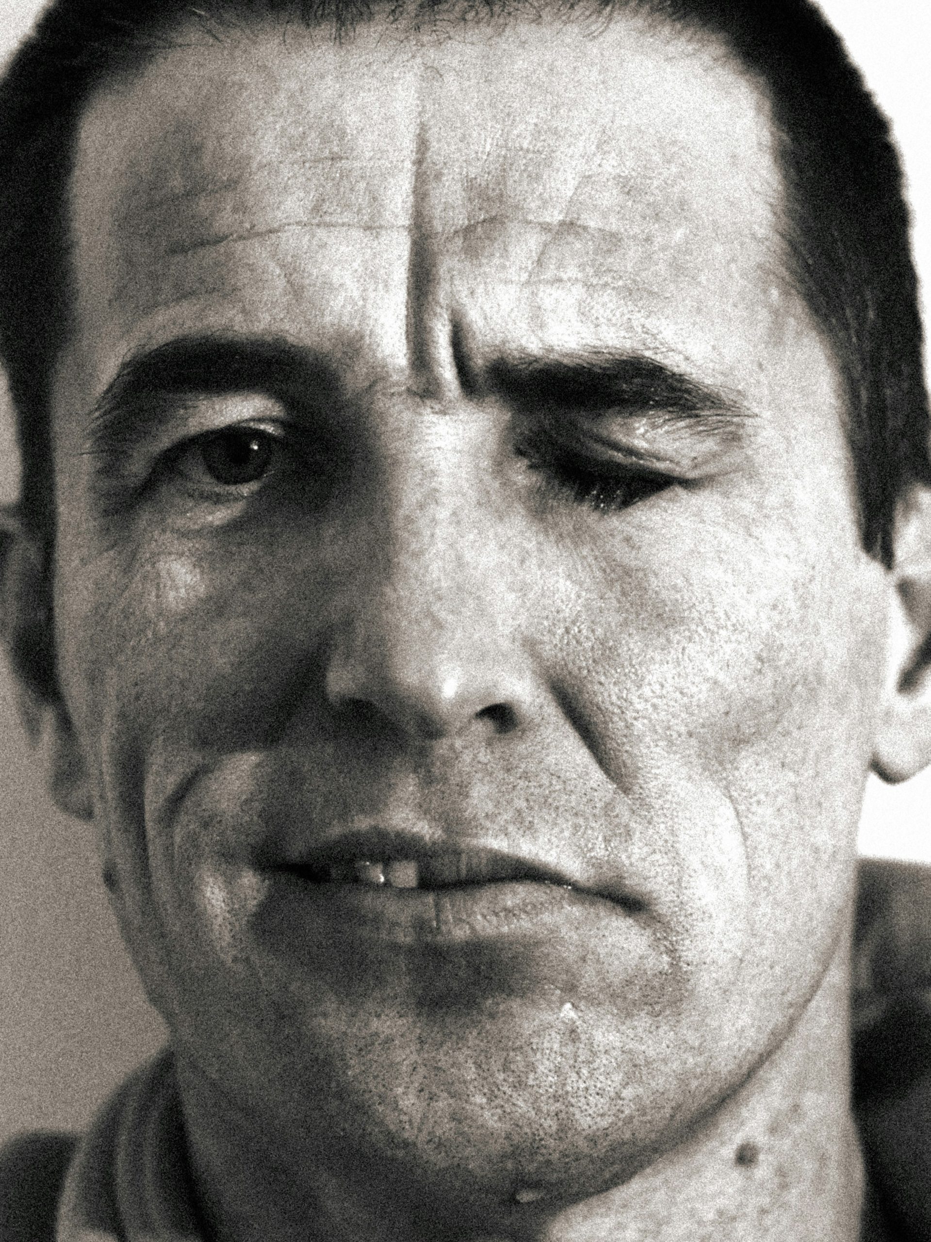
Bell’s palsy is the leading cause of sudden unilateral facial paralysis. Because of its abrupt onset, patients often fear they are experiencing an acute ischemic stroke. Classified as a peripheral lower motor neuron (LMN) facial nerve palsy (cranial nerve VII), prompt clinical recognition, rapid differentiation from central stroke, immediate initiation of oral corticosteroids, and aggressive corneal protection are critical to ensuring optimal functional recovery.
Clinical Practice Rule
Time is Tissue: Initiate high-dose oral corticosteroids within 72 hours of symptom onset to minimize facial nerve edema and compression inside the facial canal. Corticosteroid benefit declines significantly past this 72-hour window.
1. Etiology & Risk Factors
Bell’s palsy is characterized by acute inflammation, edema, and microvascular compression of the facial nerve as it traverses the narrow, rigid fallopian (facial) canal within the temporal bone. Although idiopathic by definition, viral reactivation is strongly implicated in its pathogenesis.
| Category | Associated Pathogens / Vulnerable Populations |
|---|---|
| Viral Pathogens | Herpes simplex virus type 1 (HSV-1, most common), Herpes zoster virus (HZV), Epstein-Barr virus (EBV), Cytomegalovirus (CMV), Human immunodeficiency virus (HIV). |
| Bacterial Pathogens | Borrelia burgdorferi (Lyme disease), which should be strongly considered and tested in endemic geographic regions. |
| High-Risk Patient Groups | Pregnant individuals (particularly in the third trimester or immediate postpartum period), adults with diabetes mellitus, and individuals with severe hypertension. |
2. Clinical Presentation & Normal Function Preservations
The hallmark of Bell’s palsy is rapid-onset, complete or partial weakness of all facial expression muscles on the affected side. Weakness typically progresses to maximum intensity within 48 to 72 hours.
Motor & Autonomic Manifestations
- Motor Weakness: Inability to raise the eyebrow or wrinkle the forehead, lagophthalmos (incomplete eyelid closure), flattening of the nasolabial fold, drooping mouth corner, impaired smiling, intraoral food entrapment, and involuntary cheek biting.
- Sensory & Autonomic Signs: Dysgeusia (loss of taste on the anterior two-thirds of the tongue), hyperacusis (increased sensitivity to sound due to stapedius muscle paralysis), retroauricular or mastoid pain (otalgia), ocular dryness due to reduced lacrimation, and dysesthesia around the cheek/mouth.
Sparing of Intact Neurological Functions
Because Bell’s palsy strictly affects cranial nerve VII, several motor and sensory domains remain completely uncompromised. Normal functions include: extraocular eye movements (CN III, IV, VI), jaw movement and mastication (CN V motor), swallowing and palate elevation (CN IX, X), tongue protrusion (CN XII), facial skin tactile sensation (CN V sensory), visual fields, and olfaction.
3. Diagnostic Differentiation: Bell’s Palsy vs. Stroke
The pivotal step in physical examination is evaluating upper facial muscle function—specifically the ability to wrinkle the forehead and raise the eyebrow on the weak side.
| Condition | Forehead Involvement | Associated Neurological Findings |
|---|---|---|
| Bell’s Palsy (Peripheral / LMN) |
Affected Inability to raise eyebrow or wrinkle forehead on the involved side. Entire ipsilateral hemiface is weak. |
Isolated CN VII palsy. Normal extremity motor strength, normal speech/language, intact extraocular movements, and absent central neurological deficits. |
| Acute Stroke (Central / UMN) |
Spared Forehead movement remains intact due to bilateral cortical innervation to the facial motor nucleus for upper facial muscles. |
Frequently accompanied by contralateral hemiparesis/paresthesia, dysarthria, aphasia, ataxia, visual field cuts, or altered sensorium. |
Important Clinical Mimics to Consider
- Lyme Neuroborreliosis: Suspect in endemic regions; frequently causes bilateral facial nerve palsy.
- Ramsay Hunt Syndrome (Herpes Zoster Oticus): Characterized by severe otalgia, painful vesicular eruptions in the external auditory canal or pinna, and frequent vestibulo-cochlear involvement (vertigo, hearing loss).
- Neoplasms: Parotid gland malignancies, acoustic neuromas, or facial nerve schwannomas (suspect if onset is insidious or exceeds 8 weeks without recovery).
- Infectious / Inflammatory: Otitis media, malignant otitis externa, Guillain-Barré syndrome (Miller Fisher variant), or sarcoidosis (Heerfordt syndrome).
4. Evidence-Based Treatment Protocols
| Intervention | Dosing Regimen & Details | Clinical Rationale & Evidence |
|---|---|---|
| Corticosteroids (First-Line) |
Prednisone: 50–60 mg daily for 5 days, followed by a 5-day taper (Total dose ≥450 mg). Must initiate within 72 hours of symptom onset. | AAN Level A recommendation. Reduces facial nerve inflammation and edema, significantly increasing complete recovery rates. |
| Antiviral Therapy (Adjunctive) |
Valacyclovir: 1,000 mg TID for 7 days, OR Acyclovir: 400 mg 5 times daily for 10 days. |
AAN Level C recommendation. Added only in combination with steroids (never as monotherapy) for severe presentation to reduce synkinesis. |
| Ocular Protection (Critical) |
• Daytime: Preservative-free artificial tears Q1-2H. • Nighttime: Thick lubricating ophthalmic ointment. • Protective eyewear, moisture chambers, and bedtime eyelid taping. |
Prevents exposure keratitis, corneal desiccation, ulceration, and permanent visual loss secondary to lagophthalmos. |
Prognosis & Follow-Up Requirements
Overall prognosis for idiopathic Bell’s palsy is excellent:
- Approximately 85% of patients show initial clinical recovery within 3 weeks of onset.
- Up to 70–80% achieve full baseline facial function within 3 to 6 months without long-term sequelae.
- A small percentage may experience incomplete recovery, permanent facial asymmetry, or aberrant axonal regeneration leading to synkinesis (e.g., involuntary eye closure during smiling).
Red Flag Criteria for Diagnostic Re-Evaluation
If facial weakness continues to progress beyond 7 to 10 days, shows zero clinical improvement by 6 to 8 weeks post-onset, or is accompanied by recurrent episodes, clinicians must obtain contrast-enhanced cranial MRI and electrodiagnostic testing (EMG/ENoG) to rule out underlying structural or neoplastic etiologies.
Clinical Practice Pearls
- Forehead Sparing Equals Central Origin: Forehead motion preservation strongly indicates an upper motor neuron (stroke/mass) lesion, whereas full hemifacial involvement confirms a lower motor neuron process (Bell’s palsy).
- 72-Hour Steroid Window: Always verify symptom onset timing. Initiating oral prednisone within 72 hours provides the greatest probability of complete functional restoration.
- Protect the Cornea: Motor facial recovery is worthless if the patient develops permanent corneal scarring from unmanaged lagophthalmos. Prescribe lubricants immediately at the initial visit.
