
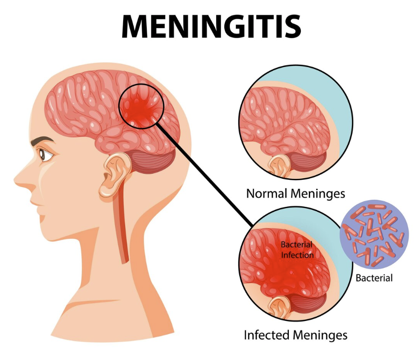
Bacterial meningitis is a neurological emergency characterized by acute infection and purulent inflammation of the leptomeninges and subarachnoid cerebrospinal fluid (CSF). Rapid clinical deterioration, permanent neurodevelopmental sequelae, septic shock, and death can occur within hours. Early recognition, prompt diagnostic evaluation, and immediate empiric antimicrobial administration are paramount to reducing mortality.
Clinical Practice Rule
Suspect early. Culture promptly. Treat immediately. Never delay empiric antimicrobial therapy to obtain neuroimaging or perform a lumbar puncture when acute bacterial meningitis is clinically suspected.
1. Pathogenesis & Microbiology
Pathogens reach the central nervous system (CNS) via three primary mechanisms:
- Hematogenous Dissemination: Most common route. Mucosal colonization (e.g., nasopharynx) leads to intravascular invasion, survival against host defenses, and crossing of the blood-brain barrier into the subarachnoid space.
- Direct Local Extension: Contiguous spread from parameningeal foci such as severe acute otitis media, bacterial sinusitis, or mastoiditis.
- Direct Inoculation: Anatomical breaches allowing pathogen entry, including basilar skull or facial fractures, CSF leaks, neurosurgical procedures, or congenital neural tube defects.
Key Bacterial Pathogens
| Organism | Microbiological Features | Clinical Relevance & Risk Factors |
|---|---|---|
| Streptococcus pneumoniae | Gram-positive diplococcus | Leading cause of bacterial meningitis in adults, elderly individuals, and immunocompromised hosts. High risk with concurrent otitis/pneumonia. |
| Neisseria meningitidis | Gram-negative diplococcus | Associated with meningococcal disease, epidemic outbreaks in young adults/dorms, and rapidly progressive purpuric rash. |
| Staphylococcus species | Gram-positive cocci (in clusters) | Associated with neurosurgical intervention, penetrating head trauma, or CSF shunts/healthcare-associated infections. |
| Haemophilus influenzae type b (Hib) | Gram-negative coccobacillus | Incidence dramatically reduced due to routine pediatric conjugate vaccination; remains a consideration in unvaccinated or asplenic patients. |
Meningococcal Disease Considerations
Neisseria meningitidis spreads via respiratory droplets during close, prolonged contact. Infectious drop-off occurs rapidly following effective antibiotic initiation. Suspected cases require strict respiratory droplet precautions, urgent public health reporting, and post-exposure chemoprophylaxis for high-risk close contacts regardless of vaccination status.
2. Clinical Presentation & Diagnostic Signs
The classical clinical triad consists of fever, severe headache, and nuchal rigidity. However, fewer than 50% of adult patients present with all three features simultaneously. Clinicians must maintain high suspicion in patients presenting with altered mental status, photophobia, confusion, or new-onset seizures.
Petechiae & Purpura Warning
A non-blanching petechial or purpuric skin rash strongly suggests invasive meningococcemia. This represents a fulminant systemic process that can rapidly evolve into Waterhouse-Friderichsen syndrome, disseminated intravascular coagulation (DIC), severe septic shock, and multiorgan failure.
Meningeal Examination Signs
- Brudzinski Sign: Passive flexion of the neck in a supine patient elicits involuntary flexion of the hips and knees.
- Kernig Sign: With the hip flexed at 90°, attempted knee extension induces severe posterior thigh pain or resistive resistance.
Clinical Pearl: Kernig and Brudzinski signs have high specificity but low sensitivity. Their absence does not exclude acute meningitis.
3. Differential Diagnosis: Meningitis vs. Encephalitis
| Feature | Meningitis (Bacterial vs. Viral) | Encephalitis |
|---|---|---|
| Primary Site | Meninges & subarachnoid space (CSF) | Brain parenchyma |
| Headache | Prominent & diffuse | Common |
| Nuchal Rigidity | Classic finding (especially in bacterial) | Often absent or mild |
| Altered Mental Status | Possible in late/severe bacterial cases | Prominent early feature (confusion, lethargy) |
| Neurological Deficits / Seizures | Less common initially | Prominent & early (focal deficits, behavioral change) |
4. Cerebrospinal Fluid (CSF) Analysis & Imaging Protocols
Lumbar puncture (LP) with CSF analysis is the definitive diagnostic procedure. Blood cultures should be drawn immediately prior to antimicrobial administration.
| CSF Parameter | Normal Values | Bacterial Meningitis | Viral (Aseptic) Meningitis |
|---|---|---|---|
| Opening Pressure | 50–200 mm H₂O | Markedly Elevated (>200–300 mm H₂O) | Normal to mildly elevated |
| WBC Count | <5 cells/mm³ | Markedly Elevated (1,000–10,000+ /mm³) | Elevated (usually 10–500 /mm³) |
| Predominant Cell Type | Mononuclear | Neutrophils (>80%) | Lymphocytes |
| Glucose Concentration | 40–70 mg/dL (>60% serum) | Markedly Decreased (<40% serum ratio) | Usually Normal |
| Protein Concentration | 15–45 mg/dL | Markedly Elevated (100–500+ mg/dL) | Normal to mildly elevated |
Indications for Neuroimaging (CT Head) Prior to LP
Head CT before LP is not required routinely and should be reserved for patients with specific risk factors for herniation or mass lesions:
- Impaired consciousness (GCS <10 or markedly decreased)
- Focal neurological deficits
- New-onset seizures within 1 week
- Papilledema on fundoscopic examination
- Immunocompromised state (e.g., HIV, active immunosuppressive therapy)
- History of central nervous system disease (mass, stroke, focal infection)
5. Management & Prevention Protocols
Empiric Treatment & Prophylaxis
- Empiric Antibiotics: Rapid parenteral administration of high-dose broad-spectrum agents (typically 3rd-generation cephalosporin such as ceftriaxone/cefotaxime PLUS vancomycin, plus ampicillin if Listeria monocytogenes is suspected in elderly or immunocompromised patients).
- Adjunctive Corticosteroids: Intravenous dexamethasone administered prior to or concurrently with the first dose of antibiotics reduces hearing loss and mortality in pneumococcal meningitis.
- Empiric Antivirals: Add intravenous acyclovir if HSV encephalitis is clinically suspected pending PCR results.
- Meningococcal Chemoprophylaxis: Indicated for close contacts exposed to N. meningitidis within 7 days prior. Options include oral Rifampin, single-dose IM Ceftriaxone, or oral Ciprofloxacin.
Prevention via Active Immunization
- Pneumococcal Vaccines: Conjugate (PCV15/PCV20) and polysaccharide (PPSV23) vaccines target invasive S. pneumoniae.
- Meningococcal Vaccines: Quadrivalent MenACWY targeting serogroups A, C, W, Y, and MenB targeting serogroup B.
- Hib Vaccine: Routine primary pediatric conjugate series.
Clinical Practice Pearls
- Antibiotics Take Priority: If neuroimaging is required prior to LP, perform blood cultures first, immediately initiate empiric antibiotics and corticosteroids, and then send the patient for CT scan.
- Hallmark CSF Triad: Neutrophilic pleocytosis + low CSF glucose (<40% serum) + elevated protein is pathognomonic for bacterial etiology until proven otherwise.
- Chemoprophylaxis Window: Post-exposure antimicrobial prophylaxis for meningococcal disease contacts should be administered as soon as possible, ideally within 24 hours of identification.
