
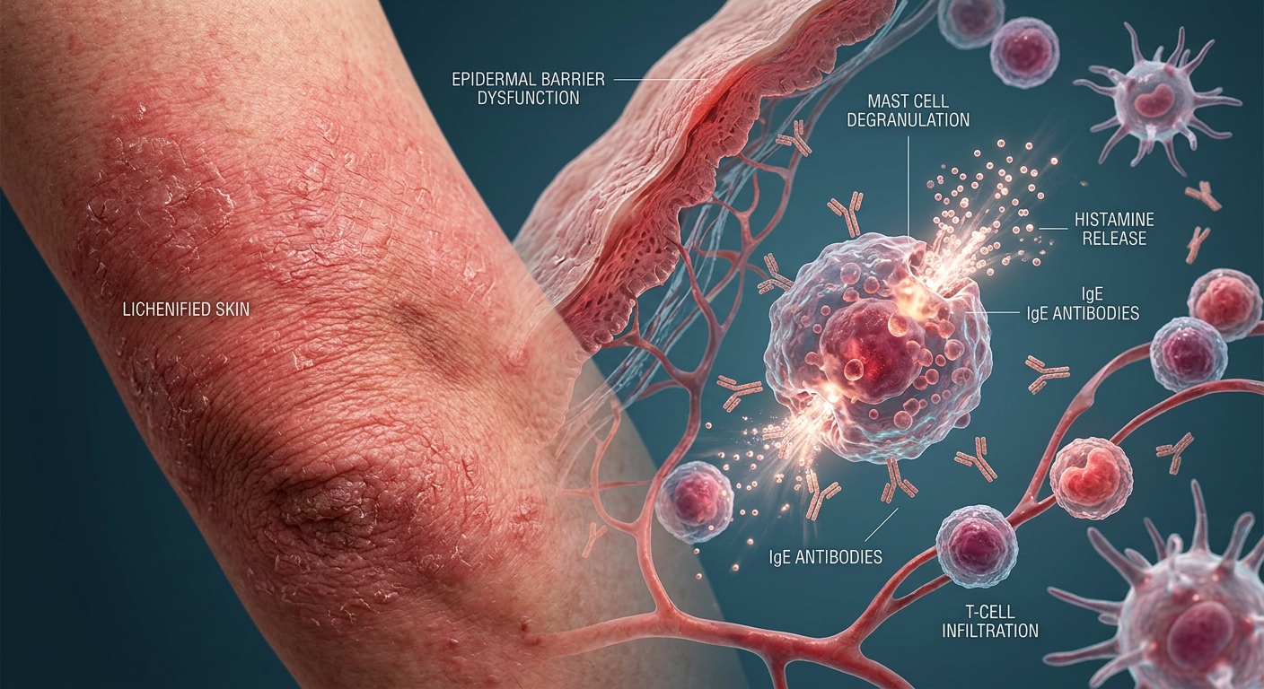
Atopic dermatitis (eczema) is a chronic, relapsing inflammatory skin disease driven by epidermal barrier dysfunction and Type I IgE-mediated hypersensitivity. Successful clinical management requires a multi-pronged approach: repairing the skin barrier, controlling acute inflammatory flares, and systematically managing nocturnal pruritus.
Pathophysiology & Diagnostic Core Principles
- Type I Hypersensitivity Cascade: Antigen exposure triggers IgE cross-linking on mast cell surfaces, releasing histamine and pro-inflammatory cytokines that drive vasodilation, edema, and intense pruritus.
- The Atopic Triad: Strongly linked by familial genetics to allergic rhinitis, asthma, and food allergies/atopic gastroenteropathy.
- Pruritus-First Criterion: Diagnosis strictly requires pruritus (itching and subsequent scratching) plus at least 3 supporting criteria: early age of onset (<5 yrs), history of dry skin/scaling, personal/family atopic history, or typical flexural rash.
- Laboratory & Culture Roles: Routine skin swab cultures yield normal cutaneous flora and are not routinely indicated unless secondary bacterial infection (e.g., S. aureus impetiginization) is clinically suspected.
1. Age-Dependent Presentation & Distribution
| Patient Demographics | Typical Lesion Distribution | Morphology & Clinical Features |
|---|---|---|
| Infants & Toddlers (<2 years old) |
Facial extensor predominance: cheeks, scalp, forehead, and neck. Diaper area is characteristically spared. | Erythematous papules, vesicles, weeping, and crusting. Diaper area is spared due to the moist, protective, occlusive barrier. |
| Older Children & Adolescents (2–12 years old) |
Flexural surfaces: antecubital fossae, popliteal fossae, wrists, and ankles. Groin and axillae spared. | Lichenification (thickened skin with exaggerated skin markings) from chronic rubbing, dry scaling, and excoriations. |
| Adults (>12 years old) |
Flexural folds, hands, eyelids, neck, and perioral region. | Severe generalized xerosis, chronic lichenified plaques, prominent hand dermatitis, and post-inflammatory pigment change. |
2. Comprehensive Step-Up Pharmacotherapy
| Therapy Class | Medications / Interventions | Clinical Indications & Practice Considerations |
|---|---|---|
| Soothing Dressings | Cool, wet compresses; Burow’s solution (Domeboro®) | Apply for 20–30 minutes to soothe acute, weeping/exudative flares and facilitate softening of crusts. |
| Topical Corticosteroids (TCS) | Low-to-Intermediate potency (e.g., Hydrocortisone 2.5%, Triamcinolone 0.1%) | First-line anti-inflammatory agent for flares. Use intermediate potency to control acute flares, then taper down to lowest effective potency. Avoid high-potency agents on face/skin folds. |
| Topical Calcineurin Inhibitors (TCI) | Pimecrolimus (Elidel®), Tacrolimus (Protopic®) | Steroid-sparing agents that block T-cell activation. Excellent for delicate skin (face, eyelids, intertriginous areas). Black Box Warning: Contraindicated in children <2 years old. |
| PDE-4 Inhibitors | Crisaborole (Eucrisa®) 2% ointment | Non-steroidal option approved for mild-to-moderate disease in patients ≥3 months old (applied BID). May cause localized application-site burning/stinging. |
| Targeted Biologics | Dupilumab (Dupixent®) SQ | Monoclonal antibody targeting IL-4Rα (inhibiting IL-4/IL-13 signaling) for moderate-to-severe refractory disease failing topical therapy. |
Pruritus Control & Oral Antihistamine Strategy
Pruritus directly impairs sleep quality and drives the “itch-scratch-cycle” that breaks down the epidermal barrier.
- Nighttime Sedating Options: First-generation sedating antihistamines—specifically Hydroxyzine (Atarax®) or Doxepin—are highly effective at bedtime to suppress nocturnal pruritus via central sedation.
- Daytime Non-Sedating Options: Active metabolites like Cetirizine (Zyrtec®) or Levocetirizine can be used for daytime symptom control or co-existing allergic rhinitis.
- Clinical Avoidance: Topical antihistamines are ineffective and strongly discouraged due to high risks of contact sensitization and allergic contact dermatitis.
3. Differential Diagnosis & Non-Pharmacologic Management
Non-Pharmacologic Barrier Repair & Rule-Outs
- Skin Barrier Optimization: Emphasize short, lukewarm showers/baths (<10 mins), use soap-free cleansers, and apply thick emollient ointments/creams (e.g., petrolatum, ceramide formulations) within 3 minutes of bathing (“soak and seal” method).
- Differential Rule-Outs: Differentiate from Lichen Simplex Chronicus (localized circumscribed plaque from habitual scratching), Nummular Eczema (coin-shaped plaques), Plaque Psoriasis (silvery scale on extensor surfaces), Scabies (burrows in web spaces), Seborrheic Dermatitis (greasy yellow scale), and Tinea Corporis (annular lesion with central clearing).
High-Yield Exam & Practice Pearls
- The “Diaper Area” Trap: Infantile eczema affects the face and scalp but spares the diaper area. If the diaper region is inflamed with satellite lesions, suspect Candida diaper dermatitis or seborrheic dermatitis.
- Age Cut-off Warning: Topical calcineurin inhibitors (Tacrolimus, Pimecrolimus) are strictly contraindicated in children under 2 years of age.
- Topical Antihistamine Mistake: Avoid prescribing topical diphenhydramine or topical antihistamines—they offer poor efficacy and frequently induce allergic contact dermatitis.
- Eczema Herpeticum Alert: Monomorphic, punched-out erosions over pre-existing eczema indicate HSV superinfection (Eczema Herpeticum)—a medical emergency requiring prompt systemic Acyclovir/Valacyclovir.
