
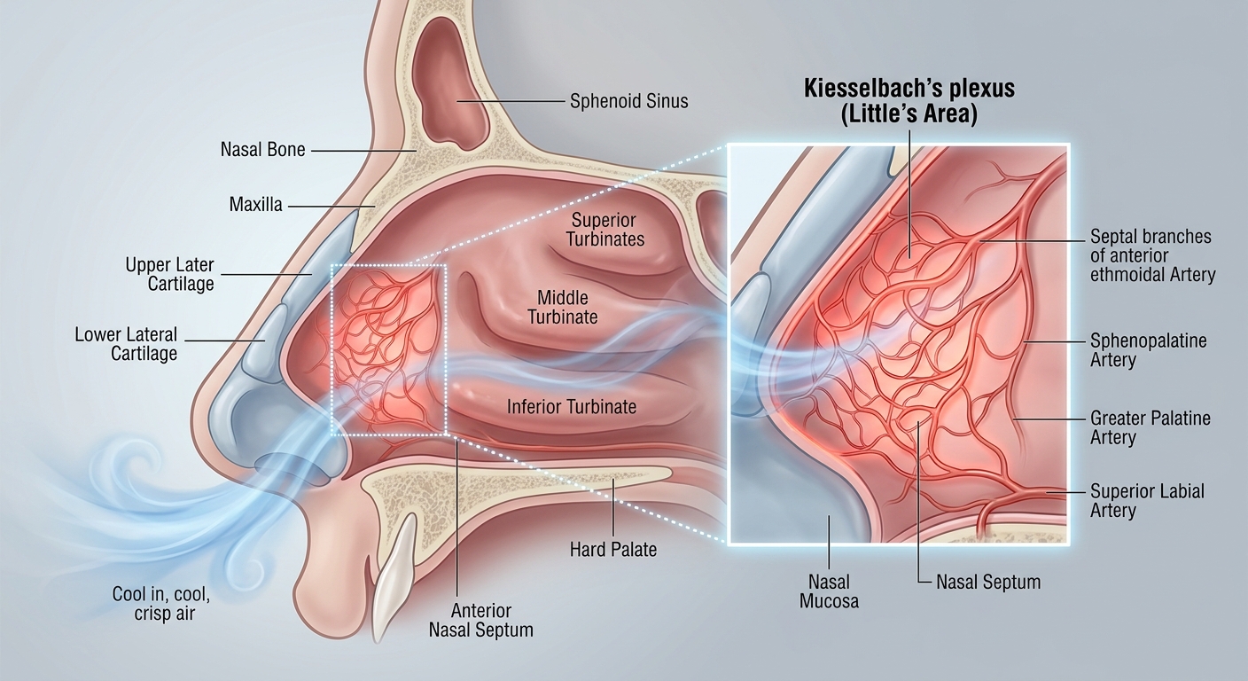
Anterior Epistaxis (commonly referred to as a nosebleed) represents an acute bleeding event originating from the anterior nasal cavity. It is the most frequent presentation of nasal bleeding encountered across acute care and outpatient settings, accounting for the vast majority of clinical epistaxis encounters.
Epidemiology, Etiology & Vascular Anatomy
- Anatomic Origin (~90% of Cases): Kiesselbach’s Plexus (Little’s Area) on the anterior nasal septum—a highly vascular confluence of the anterior ethmoidal, sphenopalatine, greater palatine, and superior labial arteries.
- Bimodal Age Distribution: Children (<10 years) and older adults (45–65 years). Slightly higher prevalence in males.
- Primary Triggers & Mechanics:
- Local Mucosal Dryness: Highly prevalent during winter months or low-humidity environments; causes mucosal fragility, crusting, and micro-fissures.
- Mechanical Microtrauma: Digital manipulation (nose picking), vigorous nose blowing, nasal instrumentation, or blunt nasal trauma.
- Clinical Clarification on Hypertension: While elevated blood pressure is frequently present during acute encounters due to anxiety and pain, there is no specific blood pressure threshold that directly triggers epistaxis.
1. Diagnostic Approach & Evaluation Strategy
| Diagnostic Pillar | Clinical Action | Rationale & Exam Pearls |
|---|---|---|
| Primary Assessment | Anterior Rhinoscopy & Direct Visualization | Anterior epistaxis is fundamentally a clinical diagnosis. Anterior bleeding sites in Kiesselbach’s plexus are readily visualizable using a speculum/penlight following gentle clearing of blood clots. |
| Laboratory Evaluation | PT/INR, PTT, CBC (Coagulation Studies) | Routine labs are NOT recommended. Obtain coagulation studies only if the patient has a history of bleeding diatheses, suspected systemic coagulopathy, heavy alcohol use, or if bleeding remains refractory to standard measures. |
| Anatomic Differentiation | Rule out Posterior Epistaxis | Suspect a posterior origin (Woodruff’s plexus) if bleeding is bilateral, flows profusely down the posterior pharynx despite anterior compression, or fails anterior packing. |
2. Stepwise Hemostatic Escalation Protocol
| Step / Level | Clinical Action & Technique | Operational Guidance & Pitfalls |
|---|---|---|
| Step 1 First-Line |
Firm Direct Compression | Pinch the soft anterior portion of the nose (superior to alar cartilage) continuously for at least 10 unbroken minutes. Head should be leaned slightly forward to prevent blood swallowing. Critical Pitfall: Avoid releasing pressure intermittently to check if bleeding has stopped, as this disrupts early fibrin clot formation. |
| Step 2 Second-Line |
Topical Agents & Nasal Packing | • Topical Therapies: Apply topical Tranexamic Acid (TXA) or topical vasoconstrictors (e.g., oxymetazoline 0.05%). • Anterior Packing: Insert absorbable (e.g., oxidized cellulose, gelatin sponge) or non-absorbable packing (e.g., nasal tampons, pre-formed sponges) to deliver direct mechanical pressure. |
| Step 3 Refractory Vessel |
Chemical / Electrical Cautery | Indicated when a single anterior vessel is clearly visualizable following clot evacuation. • Perform localized chemical cautery using silver nitrate applicators for 3–5 seconds. Safety Alert: Cauterize only one side of the septum at a time to prevent septal perforation. |
| Step 4 Emergent ENT |
Urgent ENT Consultation | Required when bleeding remains uncontrolled despite packing, topical agents, and cautery. • ENT management includes endoscopic evaluation, electrocautery, surgical ligation (e.g., sphenopalatine artery ligation), or endovascular embolization. |
3. Special Populations & High-Yield Comparisons
| Clinical Scenario | Pathophysiologic Impact | Tailored Management Strategy |
|---|---|---|
| Anticoagulated Patients (Warfarin, DOACs, Antiplatelets) |
Active anticoagulation (e.g., Rivaroxaban, Apixaban, Warfarin) impairs primary and secondary hemostasis, leading to frequent failure of conservative compression. | Expect early failure with Step 1 compression. Escalate rapidly to Step 2 (Topical TXA / Nasal Packing). Routine reversal of anticoagulation is generally not required unless life-threatening hemorrhage occurs. |
| Anterior vs. Posterior Epistaxis | Anterior = Kiesselbach’s plexus (~90%). Posterior = Woodruff’s plexus (~10%; sphenopalatine/ethmoidal branches). |
Posterior bleeds occur in older adults with vascular disease, present with profuse pharyngeal bleeding, and require posterior packing/balloon catheters or surgical intervention. |
Critical Procedural Pitfall: Septal Perforation Risk
When performing chemical cautery with silver nitrate, NEVER cauterize bilateral sides of the nasal septum simultaneously. Bilateral cauterization risks ischemic necrosis of the septal cartilage, leading to permanent septal perforation.
High-Yield Exam & Practice Pearls
- Anatomic Landmark: ~90% of all epistaxis cases originate from Kiesselbach’s plexus (Little’s area) in the anterior nasal cavity.
- Primary Causes: Local mucosal dryness (especially in cold winter air) and mechanical trauma (nose picking/digital trauma).
- Hypertension Myth: Elevated blood pressure during a nosebleed is usually secondary to anxiety; no specific BP threshold triggers epistaxis.
- Laboratory Rule: Diagnosis is clinical; routine lab work (PT/INR, CBC) is unnecessary unless bleeding is refractory or coagulopathy is suspected.
- First-Line Standard: Firm, continuous direct pressure pinching the soft anterior nose for a full 10 minutes without checking.
- Refractory Order: Compression → Topical TXA / Vasoconstrictors / Nasal Packing → Silver Nitrate Cautery → 🚨 ENT Consultation.
- Anticoagulant Shift: Patients on Warfarin or DOACs often fail simple compression and require early packing/topical TXA.
