
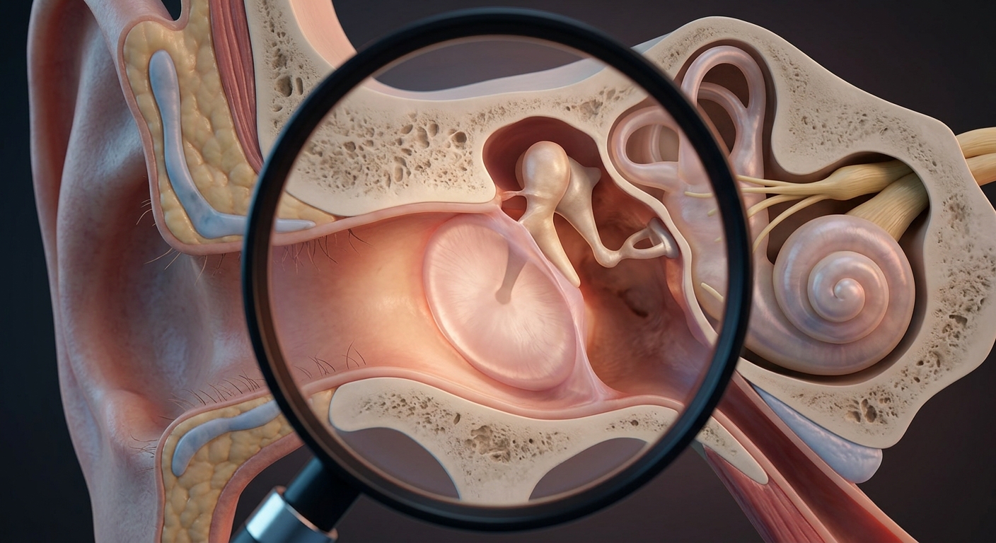
Acute Otitis Media (AOM) is an acute infectious and inflammatory process affecting the middle ear space. While classically recognized as a pediatric disease, AOM is a frequent diagnosis in adolescents and adults presenting with acute otalgia to primary care, urgent care, and emergency departments.
Pathophysiology & Microbiological Profile
- Pathophysiological Sequence:
Viral URI / Allergic Rhinitis → Eustachian Tube Dysfunction → Negative Middle-Ear Pressure → Pharyngeal Pathogen Aspiration → Acute Otitis Media - Bacterial Pathogens (S-H-M Trio):
- Streptococcus pneumoniae (Most common bacterial driver; high-dose amoxicillin target).
- Haemophilus influenzae (Nontypeable strains predominate).
- Moraxella catarrhalis (High rate of beta-lactamase production).
- Viral Etiologies: Human Rhinovirus, RSV, Adenovirus, and Influenza (purely viral cases typically self-resolve in 7–10 days with supportive care).
1. Diagnostic Strategy & Clinical Presentation
| Diagnostic Element | Key Symptoms & Otoscopic Findings | Clinical Pearls & Testing Rules |
|---|---|---|
| Core Diagnostic Formula |
• Acute Otalgia: Deep, throbbing ear pain of rapid onset. • TM Bulging & Erythema: Diffuse redness with altered or obscured bony landmarks. • Decreased TM Mobility: Confirmed visually or via pneumatic otoscopy. |
• Pneumatic otoscopy is the gold-standard bedside method to verify reduced tympanic membrane mobility. • Pain is not exacerbated by tragus press or pinna traction (key clinical separator from Otitis Externa). |
| Microbiological Testing |
• Routine tympanocentesis or throat cultures are not recommended. • Pathogens are predictable, making empiric therapy the standard of care. |
Cultures are strictly reserved for rare cases of initial treatment failure, complex cases, or severe refractory infection in immunocompromised hosts. |
2. Systemic Antimicrobial & Management Protocols
| Clinical Scenario | First-Line & Alternative Regimens | Strategic Management Considerations |
|---|---|---|
| Analgesic Support (All Patients) | Oral NSAIDs (Ibuprofen) or Acetaminophen. | Provides essential symptom relief during the first 24–48 hours until systemic antimicrobial therapy halts bacterial replication. |
| No Antibiotics in Past Month Standard Risk |
• First-Line: High-dose Amoxicillin 1,000 mg PO TID. • Oral Cephalosporins: Amoxicillin-clavulanate ER 2,000/125 mg PO BID, Cefdinir (300 mg BID or 600 mg daily), Cefpodoxime 200 mg BID, or Cefprozil. |
Adult AOM warrants prompt systemic antibiotics (unlike pediatric watchful waiting). Standard treatment duration: 5–7 days. |
| Antibiotics Used in Past Month DRSP Risk |
• First-Line: Amoxicillin-clavulanate ER 2,000/125 mg PO BID. • Parenteral Option: Ceftriaxone 1–2 g IV/IM daily for 3 days. |
Recent exposure increases the probability of Drug-Resistant S. pneumoniae (DRSP) and beta-lactamase-producing organisms. Broader coverage is required. |
| Beta-Lactam Hypersensitivity |
• Non-IgE (Mild/Rash): Cefdinir, Cefpodoxime, or Cefprozil. • IgE-Mediated (Anaphylaxis/Hives): Non-beta-lactam options (e.g., fluoroquinolones or doxycycline). |
Avoid all cephalosporins in confirmed IgE-mediated reactions. Note: Macrolides (Azithromycin) and TMP-SMX have high DRSP resistance rates. |
3. Disease Trajectory & Post-AOM Effusion (OME)
| Phase | Expected Clinical Progression | Management Protocol |
|---|---|---|
| 1–2 Weeks Post-Treatment | Resolving acute inflammation: TM mobility normalizes, erythema fades, and visible landmarks reappear. Cervical lymphadenopathy subsides. | Reassurance; no further interventions needed if pain and fever have resolved. |
| 4–6 Weeks Post-Treatment Otitis Media with Effusion (OME) |
Sterile serous middle-ear fluid persists behind an intact TM. Patients may report ear popping, cracking, fullness, or mild conductive hearing loss. | Do NOT re-treat with antibiotics. Post-AOM serous effusion is non-infectious and resolves spontaneously. Intervention is reserved for fluid persisting >3 months or significant hearing loss. |
4. Differentiating Otitis Media vs. Otitis Externa
| Feature | Acute Otitis Media (AOM) | Otitis Externa (OE) |
|---|---|---|
| Anatomic Site | Middle ear cavity (behind TM). | External auditory canal ± pinna. |
| Preceding Factor | Viral URI, allergic rhinitis, Eustachian tube dysfunction. | Swimming, moisture, cotton swab microtrauma. |
| Tragus / Pinna Sign | Negative (No pain on movement). | Positive (Severe pain on movement). ⭐ |
| Otoscopy | TM erythematous, bulging, poorly mobile; canal clear. | Canal swollen, edematous, debris-filled; TM often normal. |
| Pathogens & Tx | S. pneumoniae, H. influenzae, M. catarrhalis → Systemic Antibiotics. | P. aeruginosa, S. aureus → Topical Drops. |
Bedside Diagnostic Shortcut
Press the tragus or pull the pinna: Pain present? → Think Otitis Externa. Pain absent + abnormal TM post-URI? → Think Acute Otitis Media.
High-Yield Exam & Practice Pearls
- Pathopathogenic Core: Antecedent URI or allergic rhinitis leads to Eustachian tube dysfunction and negative pressure in the middle ear.
- Bacterial Trio: S. pneumoniae, H. influenzae, and M. catarrhalis (S-H-M).
- Diagnostic Pairing: Severe Otalgia + Bulging/Poorly Mobile TM = Acute Otitis Media.
- Adult Strategy: Always treat adult AOM with systemic oral antibiotics (5–7 day course); do not apply pediatric “watchful waiting.”
- Resistance Rule: Antibiotics used within 30 days = Risk for Drug-Resistant S. pneumoniae (DRSP) → Use high-dose Augmentin ER or Ceftriaxone.
- Post-AOM Effusion (OME): Serous fluid lingering 4–6 weeks post-AOM is expected; do not give a second course of antibiotics for asymptomatic OME.
