
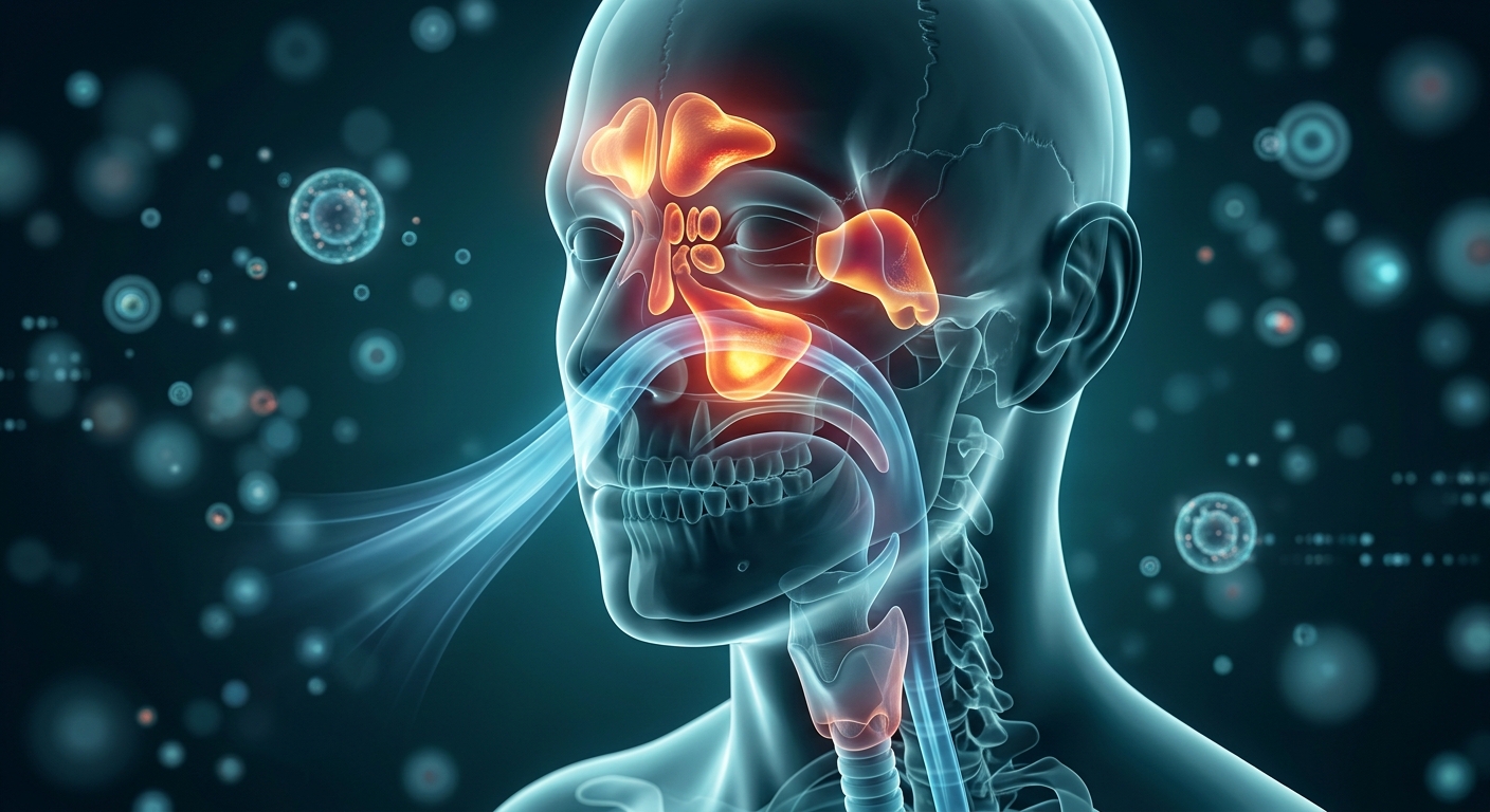
Acute Bacterial Rhinosinusitis (ABRS) is an inflammation of the paranasal sinus mucosa caused by secondary bacterial proliferation following impaired sinonasal drainage. Because viral upper respiratory infections (URIs) present with nearly identical symptoms, the cornerstone of clinical management is accurate identification of bacterial disease using specific temporal patterns, avoiding unnecessary antibiotic use and preventing treatment failure.
Pathophysiology, Predisposing Factors & Microbial Profile
- Pathophysiologic Cascade: Impaired Sinus Drainage → Retained Mucous Secretions → Stagnant Nidus → Bacterial Proliferation → Mucosal Inflammation.
- Key Risk Factors: Viral URIs (edema & ciliary loss), allergic rhinitis, tobacco smoke/environmental irritants, and anatomical blocks (septal deviation, polyps).
- Microbial Triad (S-H-M):
- Streptococcus pneumoniae: #1 pathogen (~25% resistant via altered PBPs).
- Haemophilus influenzae: #2 pathogen (30–40% resistant via β-lactamase).
- Moraxella catarrhalis: #3 pathogen (~90% resistant via β-lactamase).
1. Diagnostic Criteria & Clinical Presentations
| Diagnostic Pattern | Clinical Criteria & Trajectory | Key Differentiation vs. Viral URI |
|---|---|---|
| 1. Persistent Symptoms (P) | Nasal discharge (purulent or clear) OR facial pain/pressure lasting ≥10 days without evidence of clinical improvement. | Viral URIs typically peak by day 3–5 and resolve completely within 7–10 days. |
| 2. Severe Symptoms (S) | High fever (≥102°F / 39°C) accompanied by purulent discharge OR severe facial pain for ≥3–4 consecutive days at onset. | Viral URIs rarely produce prolonged high fevers with severe localized pain early on. |
| 3. “Double Sickening” (D) | Initial mild improvement of a typical viral URI (3–4 days), followed by sudden acute worsening with new fever, discharge, or headache. | Reflects secondary bacterial superinfection following primary viral mucosal damage. |
2. Imaging Guidelines & Differential Diagnosis
| Category | Acute Bacterial Rhinosinusitis (ABRS) | Allergic Rhinitis |
|---|---|---|
| Dominant Symptoms | Facial pain/pressure, systemic malaise, persistent nasal congestion. | Nasal/ocular pruritus, repetitive sneezing, clear rhinorrhea. |
| Nasal Secretions | Thick, opaque, or purulent (neutrophil influx). | Clear, watery, profuse. |
| Systemic Features | Fever possible, prominent facial pressure. | Fever consistently absent; allergic shimmers/salute. |
| Role of Imaging | Routine CT/X-rays NOT recommended. Reserved strictly for orbital/intracranial complications, treatment failure, or recurrent refractory cases (≥4/year). | |
Evaluation for Drug-Resistant S. pneumoniae (DRSP) & High-Risk Drivers
Assess for resistance risks prior to prescribing empiric antimicrobials:
- Age Extremes: Patients <2 years or >65 years.
- Recent Antimicrobial Exposure: Systemic antibiotic usage within the preceding 30 days.
- Recent Hospitalization: Inpatient stay within the prior 5 days.
- Comorbidities & Environment: Immunocompromised state, chronic heart/lung/liver disease, or day care attendance.
3. Pharmacotherapy & Antimicrobial Regimens
| Clinical Scenario | Preferred First-Line Regimen | Alternative / β-Lactam Allergy Options |
|---|---|---|
| Standard / Low-Risk Adult | Amoxicillin-Clavulanate 2,000 mg / 125 mg PO BID (High-dose amoxicillin overcomes DRSP; Clavulanate neutralizes β-lactamases). |
Doxycycline 100 mg PO BID (or 200 mg PO daily) OR Respiratory Fluoroquinolone (Levofloxacin/Moxifloxacin). |
| High Resistance Risk / 1st-Line Failure | High-Dose Amoxicillin-Clavulanate OR 2nd/3rd Gen Cephalosporins (Cefpodoxime, Cefprozil, Cefdinir). | Levofloxacin 750 mg PO daily OR Moxifloxacin 400 mg PO daily. |
| Contraindicated Class | DO NOT USE MACROLIDES (Azithromycin, Clarithromycin) due to >30–40% S. pneumoniae resistance. | |
High-Yield Exam & Practice Pearls
- P-S-D Rule: Diagnose ABRS strictly via Persistent (≥10d), Severe (≥3–4d), or Double Sickening patterns.
- Purulence Fallacy: Green/yellow nasal discharge occurs in viral URIs due to neutrophilic influx; it is not diagnostic of bacterial disease alone.
- Top Pathogen: Streptococcus pneumoniae is the #1 cause of both ABRS and Acute Otitis Media (AOM).
- β-Lactamase Coverage: Clavulanate must be added to amoxicillin to cover β-lactamase-producing H. influenzae and M. catarrhalis.
- No Macrolides: Avoid Azithromycin due to widespread pneumococcal resistance and high failure rates.
- No Routine CT: Imaging is unnecessary for uncomplicated cases and fails to reliably differentiate viral from bacterial sinus inflammation.
- Adjunctive Winners: Recommend nasal saline irrigation and intranasal corticosteroids; avoid routine decongestants (risk of rhinitis medicamentosa >3 days).
- Reassessment Timeline: Re-evaluate any patient failing to demonstrate clinical improvement after 3–5 days of empiric therapy.
